Download
1 / 45
490 likes | 758 Views
Bacterial Vascular Wilts. Today we will discuss bacterial vascular wilts. Vascular wilts. Erwinia tracheiphila. M. Daughtrey. R. X. Latin. Xanthomonas campestris pv . pelargonii. Pathogen detection. Bacterial wilts affect mostly herbaceous plants including vegetables and ornamentals.

E N D
Today we will discuss bacterial vascular wilts Vascular wilts Erwiniatracheiphila M. Daughtrey R. X. Latin. Xanthomonascampestrispv. pelargonii Pathogen detection
Bacterial wilts affect mostly herbaceous plants including vegetables and ornamentals
Vascular wilts are caused by bacteria that infect and block xylem vessels of host plants Healthy xylem vessels Infected xylem vessel Interfere with translocation of water and nutrients
Wilt bacteria often destroy (dissolve) parts of cell walls of xylem vessels Xylem vessel wall dissolution
Bacteria spread into intercellular spaces and can ooze out of stomata onto the leaf surface Intercellular space Bacteria Goodman and white, 1981
Bacterial Blight is the most destructive disease of geranium (Pelargoniispp.) Xanthomonascampestrispv. pelargonii
X. campestrispv.pelargoniiis a host specific pathogen that only infects Geranium PathovarTerm used for bacteria this is equivalent to the term forma specialis for fungi • 125 known pathovars of X. campestris! Defined by host range
X. campestrispv. pelargoniiis a rod shaped bacterium with single polar flagella Single polar flagellum • Rod shaped
X. campestrispv. pelargoniisurvive as an epiphyte or a saprophyte • When conditions are unfavorable for disease, the bacteria survives on plant surfaces • The bacteria can survive for up to 6 months in soil Epiphyte • Saprophyte • Epiphyte Endophyte
Development of Geranium blight symptoms is influenced by the factors in the disease triangle Host: Geranium Pathogen: Xanthomonascampestris Environmental Factors
X. campestrispvpelargoniicauses wilting symptoms on Geranium Wilting and chlorotic or necrotic lesions Wilted leaves but petiole still turgid
Symptoms are associated with water splashing from overhead irrigation or wind driven rain Associated with hydothodes V-shaped chlorotic areas on leaf limited by leaf veins
Bacterial ooze from infected stem at vascular bundles is a diagnostic sign Bacterial ooze from vascular bundles Bacterial streaming from an infected leaf
X. campestriscan spread both long and short distances • Long distance: commercial transport • Infected rooted cuttings and plants • latent infections may not be obvious Short distance: in greenhouse • Irrigation water • Ebb & flow benches • Infested cutting tools
X. campestrisinfects through wounds and natural openings Wounds may be created from cultivation or insects 1 3 2 4 University of Sydney Infection via hydathodes
X. campestrisis host tissue specific and can spread both intracellularly and intercellularly Late infection: intercellular cortex or mesophyl Early infection: Intracellular In the xylem systemic spread
There are four mechanisms of pathogenesis inducing wilt symptoms Blocking of xylem by pathogen • Bacterial cells and polysaccharides • Inhibits transpiration of water • Polysaccharides • toxic to plant cells • Also act as blocking agents in xylem
Toxins: bacterial metabolites • Affect cell membranes • Affect turgor pressure • Tyloses: Initiated by host • Evaginated parenchyma protoplasts • Protrude into empty xylem element
Control of bacterial blight relies on prevention detection, sanitation and eradication
Establish a unidirectional movement of plants to prevent disease Elite nuclear stock Indexing Select plants Nuclear stock Renew annually Continued indexing Separate and protect Propagation stock
Indexing is a procedure used to detect the presence of particular target fungi and bacteria - + R. Wick ELISA and PCR indexing Culture indexing
Limited host resistance and effective chemical are challenges to bacterial blight control Latent infections difficult to detect Little Resistance Host: Geranium Pathogen: Xanthomonascampestris Environmental Factors overhead Irrigation long periods of rain high humidity warm temperatures No effective chemical controls
During today's lab we will use the AgdiaImmunostrip test for X. campestrisp.v.pelargonii
Agdia: Immunostrip Test for X. campestris p.v. pelargonii Y Goat anti-rabbit antibody = A strip of anti-rabbit antibody produced in goats is bound to the nitrocellulose. Rabbit anti-X. c. p. antibody only = Y A strip of anti-X.campestris pv. pelargonii made in rabbit is bound to the nitrocellulose All proteins bind irreversibly to untreated nitrocellulose, including antibodies. Once bound, cannot be removed. Bound proteins will adhere to nitrocellulose through repeated washes. 1
Agdia: Immunostrip Test for X. campestris p.v. pelargonii Goat anti rabbit antibody = Y Soak dried nitrocellulose in 5% Powdered milk solution and/or 2% bovine serum albumen Proteins stick to all available binding sites remaining on nitrocellulose (gray area) Rabbit anti X. c. p. antibody only = Y All remaining protein binding sites are blocked by addition of powdered milk protein or bovine serum albumen proteins. No additional proteins can attach to the nitrocellulose. 2
dye Y Y Y Y Y Y Y Y Y Y Agdia: Immunostrip Test for X. campestris p.v. pelargonii A small reservoir of rabbit anti-X. campestris antibody-conjugated to a nonsoluble visible dye molecule is attached to bottom of nitrocellulose strip. This complex cannot attached to the nitrocellulose because NC previously blocked. Antibody-dye complex remains soluble. Goat anti rabbit antibody Rabbit anti X. c. p. antibody Rabbit antibody with dye attached = 3
Agdia: Immunostrip Test for X. campestris p.v. pelargonii Plant tissue to test (petioles or midribs) is added to bag Sample-buffer is added to bag and tissue is homogenized. Nitrocellulose “dip-stick” is then immersed up to indicated level in the sample using the window along right side of bag. Strip is allowed to incubate in bag for 30-60 minutes. Goat anti-rabbit antibody Rabbit anti-X. c. p. antibody BACTERIAL PROTEINS Rabbit anti-Xcp with dye attached liquid with bacterial proteins migrates upwards by capillary action 4
dye Y Y Y Y Y Y Y Y Y Y Agdia: Immunostrip Test for X. campestris p.v. pelargonii As plant sample diffuses upwards and encounters antibody-dye complex, the antibody-dye complex will bind specifically to antibody-recognized bacterial proteins. This new aggregate consisting of bacterial protein antigens, antibody, and dye will continue migrating upward until it passes the layer of bound rabbit antibody which also recognizes and binds the bacterial antigen protein. BACTERIAL PROTEINS BOUND TO ANTIBODY- DYE COMPLEX Rabbit antibody with dye attached 5 BACTERIAL PROTEINS
Y Y Y Y dye Y Y Y Y Y Y Y Y Y Y Y Agdia: Immunostrip Test for X. campestris p.v. pelargonii As antigen-antibody-dye complexes pass the bound-antibody, the bound-antibody attaches to exposed antigen on the complexes stopping diffusion of that complex. Dye accumulates at site of complex binding forming visible strip of blue. Rabbit anti-Xcp antibody Bacterial protein antigens Rabbit anti-Xcp antibody Dye accumulates here only if bacterial antigen is present in sample Rabbit antibody with dye attached 6 BACTERIAL PROTEINS
Y Y Y Y Y Y dye Y Y Y Y Y Rabbit anti-X.c.p. antibody Y Y Y Y Y Y Y Y Goat anti-rabbit antibody Y Y Y Agdia: Immunostrip Test for X. campestris p.v. pelargonii Antibody-dye complexes that have not previously attached to bacterial proteins continue migrating upward. Eventually The rabbit antibody-dye complex reaches the layer of bound Goat anti-rabbit antibody. The rabbit antibody binds to the Goat anti-rabbit antibody trapping and concentrating the dye At the strip of goat antibody. This indicates that the rabbit antibody-dye complex diffused upward and that the test is working properly (negative control). Rabbit antibody with dye attached 6 BACTERIAL PROTEINS
Test Control (test good) Sample test - / + (-) (+) Test results for X. campestris detection Agdia: Immunostrip Test for X. campestris p.v. pelargonii 6
Bacterial Wilt of Cucurbits is caused by Erwiniatracheiphila Cucumber Muskmelon Squash Pumpkin Cantaloupe
Development of cucumber wilt symptoms is influenced by the factors in the disease triangle Environment conducive for disease Erwiniatracheiphila Vector
E. tracheiphilais transmitted by two main insect vectors Spotted Cucumber beetle Diabrotica StripedCucumber Beetle Acalymma
Beetles can cause feeding damage and create wounds for bacteria to enter Cotyledon damage Blossom damage Mature leaf damage
Erwiniatracheiphilacauses wilting and necrosis of cucurbits Wilting of individual leaves and petiole Interveinalchlorosis and necrosis • Eventual wilt and death of entire stem
Erwiniatracheiphilareduces fruit quality and storability • Small dehydrated fruits, wrinkled and poor taste • Infected fruits may also rot in storage • Looks like drought symptoms, except may be only on specific stems or plants at random
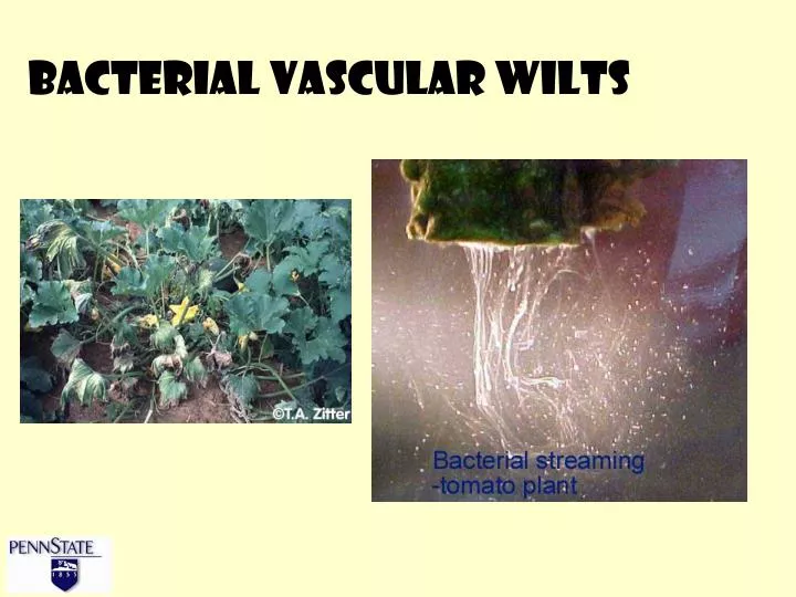
Strands of bacterial ooze on cut stem ends is a diagnostic sign of bacterial wilt Bacterial slime string
E. tracheiphilacauses symptoms by blocking vessels with polysaccharides and toxin activity Vascular bundle with bacterial ooze Erwiniatracheiphilain the lumen of a vessel element F. Gildow
Bacteria cause formation of Tyloses in the vessel elements of infected plants Lumen of vessel element Pits in wall of vessel element Layer of xylem parenchyma cells surrounding vessel elements Hypertrophiedparenchyma cell pushed through pit in vessel wall and ballooning into lumen Fig. 3-5. Agrios, 5’th ed. Pg. 112
Bacterial Wilt control relies on targeting several parts of the disease triangle Elimination of beetles – low threshold of 1 • Pesticides- reduce primary and secondary spread Remove and destroy any infected plants • Reduce secondary spread Control cucurbit weeds near field • Not many, mostly from beetles! Select resistant cultivars • Good luck – not many resistant cultivars!