Download
1 / 32
320 likes | 608 Views
4.2 Meiosis. State that meiosis is a reduction division of a diploid nucleus to form a haploid nulcei . Define homologous chromosomes Outline the process of meiosis, including pairing of homologous chromosomes and crossing over, followed by two divisions, which reults in four haploid cells.

E N D
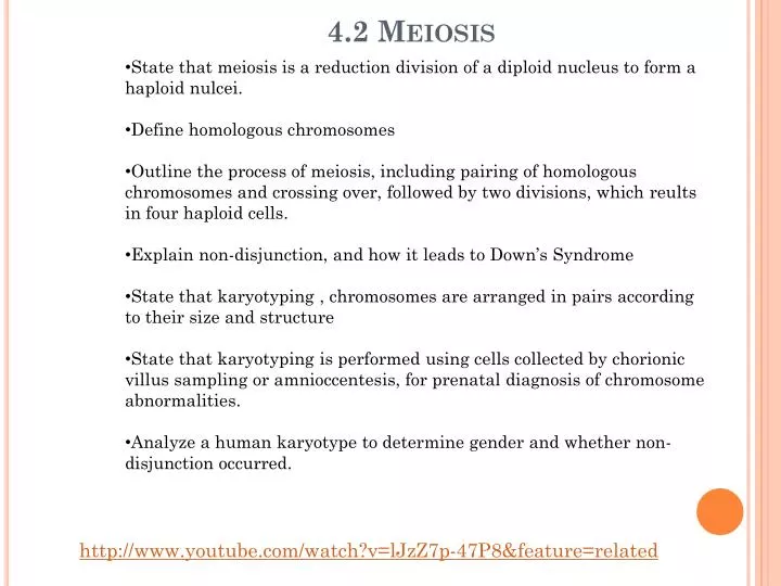
4.2 Meiosis • State that meiosis is a reduction division of a diploid nucleus to form a haploid nulcei. • Define homologous chromosomes • Outline the process of meiosis, including pairing of homologous chromosomes and crossing over, followed by two divisions, which reults in four haploid cells. • Explain non-disjunction, and how it leads to Down’s Syndrome • State that karyotyping , chromosomes are arranged in pairs according to their size and structure • State that karyotyping is performed using cells collected by chorionic villus sampling or amnioccentesis, for prenatal diagnosis of chromosome abnormalities. • Analyze a human karyotype to determine gender and whether non-disjunction occurred. http://www.youtube.com/watch?v=lJzZ7p-47P8&feature=related
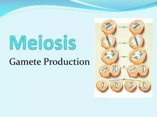
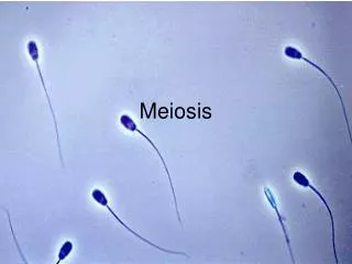
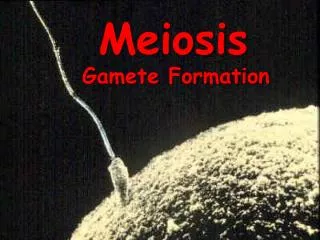
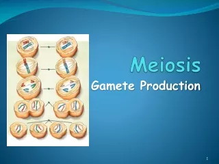
Meiosis is a type of cell division that produces gametes (sex cells). In animals they are known as sperm and egg. Each cell that is produced as a result of meiosis has half the number of chromosomes of other cells in the body.
Eukaryotic body cells have a diploid nucleus, which contains two copies of each chromosome, in homologous pairs. Homologous chromosomes- a pair of chromosomes with the same genes but not necessarily the same alleles of those genes. Humans have a diploid number of 46 chromosomes in 23 pairs, mangos have 40 chromosomes in 20 pairs, and camels have 70 in 35 pairs.
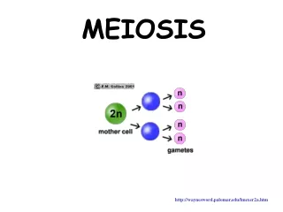
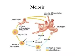
During sexual reproduction, two gametes (sex cells) fuse together so, in order to keep the chromosome number correct in the offspring, each gamete must contain only one of each chromosome pair. It must contain half the diploid number of chromosomes, which is called haploid. During gamete formation, meiosis reduces the diploid number to the haploid number. At the moment of fertilization, the normal diploid number is restored as the gametes fuse.
In humans, the haploid sperm (23 chromosomes) and the haploid egg (23 chromosomes) fuse at fertilization to form the diploid zygote, with 46 chromosomes (23 pairs).
Chromosomes Homologous Chromosomes Chromatid
The Process of Meiosis http://www.youtube.com/watch?v=kVMb4Js99tA Meiosis occurs in a series of stages, which results in the production of four cells. Mitosis, used to replace or repair cells, which is done by one cell division, meiosis involves two divisions. The first reduces the number of chromosomes by half and the second produces four gametes each containing the haploid number of chromosomes. The first division is very similar to mitosis and the second division is exactly the same as mitosis. http://www.youtube.com/watch?v=iCL6d0OwKt8
Meiosis Meiosis begins with one diploid cell containing two copies of each chromosome (one from the organism's mother and one from its father) and produces four haploid cells containing one copy of each chromosome. Each of the resulting chromosomes in the gamete cells is a unique mixture of maternal and paternal DNA, ensuring that offspring are genetically distinct from either parent. This gives rise to genetic diversity in sexually reproducing populations, which provides the variation of physical and behavioral attributes upon which natural selection acts.
Meiosis I Meiosis I separates homologous chromosomes, producing two haploid cells (23 chromosomes in humans), so meiosis I is referred to as a reductional division. A regular diploid human cell contains 46 chromosomes and it contains 23 pairs of homologous chromosomes. However, after meiosis I, although the cell contains 46 chromatids, it is only has 23 chromosomes. This is because later, in Anaphase I, the sister chromatids will remain together as the spindle fibres pull the pair toward the pole of the new cell.
Prophase I The chromosomes, which have replicated during interphase, now supercoil. Each one consists of two sister chromatids joined by the centromere. Although the genes carried by each chromosome pair are identical, the alleles may not be.
Recombination Prophase I Sister chromatids may become entangled, break and rejoin so that alleles are exchanged between them during this process called crossing over. New combinations of alleles are formed and genetic variety in the resulting gametes increases.
Prophase I The final step in the prophase I is the formation of spindle microtubles and the breakdown of the nuclear envelope.
Metaphase I Chromosomes line up on the equator at the center of the cell. Each one attached by a centromere to the spindle microtubules. The alignment of the chromosomes is random so that maternal and paternal chromosomes can appear on either side of one another on the equator. This also increases the genetic variety in the gametes.
Anaphase I The microtubules now contract towards opposite poles. The pair of sister chromatids remain together but homologous pairs are separated. This is the reduction division where the chromosome number is halved from diploid to haploid.
Telophase I Now spindle s break down and a new nuclear envelope forms. Cytokinesis follows and the cell splits into two cells, each containing only one chromosome of each homologous pair. Each chromosome, however, still consists of two sister chromatids at this point. The second division (Meiosis II will separate the two sister chromatids.
Meiosis II Meiosis II is the second part of the process. Mechanically, the process is similar to mitosis, though its genetic results are fundamentally different. The end result is production of four haploid cells (23 chromosomes, in humans) from the two haploid cells (23 chromosomes, each of the chromosomes consisting of two sister chromatids) produced in meiosis I. The four main steps of Meiosis II are: Prophase II, Metaphase II, Anaphase II, and Telophase II
Prophase II In each of the two cells resulting in Meiosis I, new spindle fibers start to form, the chromosomes recoil and the nuclear envelope begins to break down.
Metaphase II The nuclear envelope is broken down and the individual chromosomes line up on the equator of each cell. Spindle fibers from opposite ends of the cell attach to each chromatid at the centromere.
Anaphase II Sister chromatids are separated as the centromere splits and the spindle fibers pull the chromatids to opposite ends of the cell.
Telophase II Nuclear envelopes form around the four new haploid nuclei and the chromosomes now uncoil. A second cytokinesis occurs, resulting in four cells.
Non-disjunction Non-disjunction is a failure of the homologous pairs of chromosomes to separate properly during meiosis.
Non-disjunction results in gametes that contain either one too few or one too many chromosomes. Those with too few seldom survive , but in some cases a gamete with an extra chromosome does survive, which produces a zygote with three chromosomes of one type, known as trisomy.
Trisomy in chromosome 21 results in Down’s Syndrome. A gamete, usually the female one, receives 24 chromosomes instead of 23 and a baby with 47 instead of the usual 46 chromosomes in each cell.
Down’s Syndrome The incidence of Down syndrome is estimated at 1 per 733 births, although it is statistically more common with older parents due to increased mutagenic exposures upon some older parents' reproductive cells.
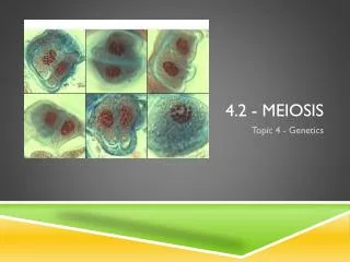
Karyotyping Chromosomes have unique banding patterns that are revealed if they are stained with specific dyes during prophase. Each chromosome has a characteristic length and has its centromere at a fixed place, and each one has a homologous partner
In a karyogram, chromosomes are stained and photographed. The image is then manipulated to arrange the chromosomes in order of their size. Karyograms indicate the sex of an individual (X,Y) Also used in prenatal diagnosis to check for chromosome abnormalities.
In the procedure called karyotyping, cells from an unborn child are collected in one of two ways: chorionic villus sampling (CVS) or amniocentesis. The cells are grown in the lab and a karyogram is prepared. This is checked for extra or missing chromosomes. The procedures are used when there is concern about potential chromosome abnormalities, such as a mother over 35 since down’s Syndrome is more common in babies of older mother.
CVS Involves taking a sample of cells from the chorionic villi, which are fine projections of the placenta embedded in the lining of the uterus. This is done 8-10 weeks into pregnancy.
Amniocentesis Takes a sample of amniotic fluid from the mother between 14 and 16 weeks for t he pregnancy
Both methods carry a small risk of damaging the fetus or even causing miscarriage. Once the results are known, the parents may choose to terminate the pregnancy if abnormalities are discovered. The tests may reveal an abnormality but cannot give indication about the severity.
Thoughts to Consider Who should make the decision to carry out the procedure? The parents or the health care officials? How important are legal and religious arguments? Both procedures carry risks of miscarriage. How can this potential risk to the unborn child be balanced with the parents’ desire for information? Does the information that can be obtained from the karyogram outweigh the risk to the unborn child? If the karyogram indicates a genetic abnormality, should the parents be permitted to consider a termination of the pregnancy?