Download
1 / 38
420 likes | 1.05k Views
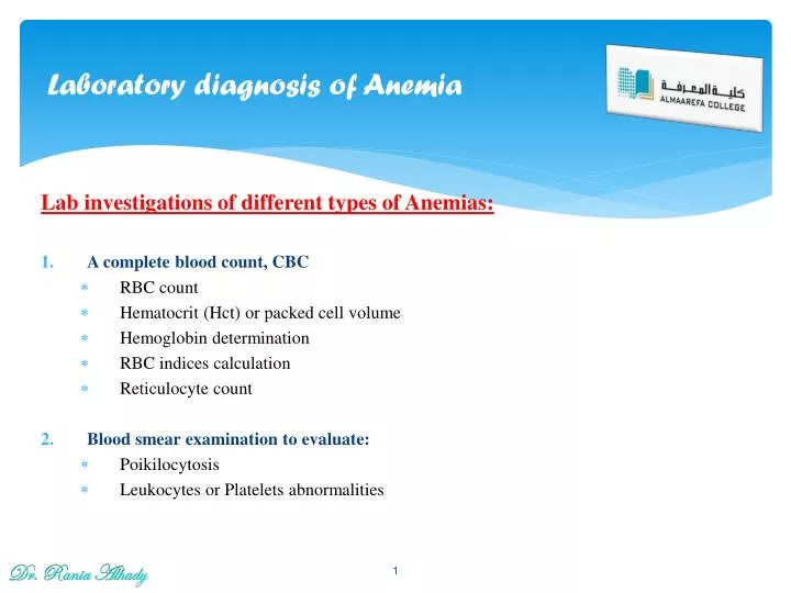
Laboratory diagnosis of Anemia. Lab investigations of different types of Anemias: A complete blood count, CBC RBC count Hematocrit (Hct) or packed cell volume Hemoglobin determination RBC indices calculation Reticulocyte count Blood smear examination to evaluate: Poikilocytosis

E N D
Laboratory diagnosis of Anemia Lab investigations of different types of Anemias: • A complete blood count, CBC • RBC count • Hematocrit (Hct) or packed cell volume • Hemoglobin determination • RBC indices calculation • Reticulocyte count • Blood smear examination to evaluate: • Poikilocytosis • Leukocytes or Platelets abnormalities Dr. Rania Alhady
Laboratory diagnosis of Anemia • A bone marrow smear and biopsy to observe: • Maturation of RBC and WBC series • Presence of megakaryocytes • Ratio of myeloid to erythroid series • Presence or absence of granulomas or tumor cells • Hemoglobin electrophoresis • Antiglobulin testing • Osmotic fragility test Dr. Rania Alhady
Red blood cell indices RBCs Indices: They are part of the complete blood count (CBC) test that provide information about the hemoglobin content and size of red blood. They are used to help diagnose the cause of anemia. The indices include: • Mean corpuscular volume (MCV): is the average size of a red blood cell and is calculated by dividing the hematocrit (Hct) by the red blood cell count. MCV = Hct / RBC Normal range: 80-100 fL(femto- is 10-15) • Mean corpuscular hemoglobin (MCH): is the average amount of hemoglobin (Hb) per red blood cell and is calculated by dividing the hemoglobin by the red blood cell count. MCH = Hb / RBC Normal range: 27-31 pg/cell(pico- is 10-12) • Mean corpuscular hemoglobin concentration (MCHC): is the average concentration of hemoglobin per red blood cell and is calculated by dividing the hemoglobin by the hematocrit. MCHC = Hb / Hct Normal range: 31-35 g/dL(deci is 10-1) Dr. Rania Alhady
Classification of Anemia Morphological classification of anemia: • Based on RBC morphology • Anemia is divided into three groups mainly on the basis of the MCV (RBC indices) • Normocytic Normochromic anemia: (normal red cell indices) • Blood loss anemia (Acute bleeding) • Hemolytic anemia (except thalassaemia) • Aplastic anemia • Pure red cell aplasia • Renal insufficciency • Anemia of endocrine disease • Toxic depression of bone marrow Dr. Rania Alhady
Classification of Anemia 2. Microcytic hypochromic anemia: ( low red cell indices) • Iron deficiency anemia • Sideroblastic anemia • Lead poisoning • Thalassemia • Chronic diseases 3. Macrocytic Normochromic ( high MCV and MCH, normal MCHC) • Megaloblastic anemia (Vit. B12 deficiency & Folic acid deficiency). • Liver disease • Post splenectomy • Hypothyroidism Dr. Rania Alhady
Classification of Anemia Dr. Rania Alhady
Microcytic- Hypochromic Anemia 1- Microcytic- Hypochromic Anemia Dr. Rania Alhady
Microcytic- Hypochromic Anemia Dr. Rania Alhady
Microcytic- Hypochromic Anemia A- Iron Deficiency Anemia (IDA) : • Is a condition in which the total body iron content is decreased below a normal level • This results in a reduced red blood cell and hemoglobin production • More than half of all anemias are due to iron deficiency. Clinical Picture: Symptoms eg. fatigue, dizziness, headache Signs eg. • Pallor • Tongue atrophy/ glossitis - raw and sore • Angular cheilosis (Stomatitis) • Spoon‑shaped nails (koilonychia), brittle nails and hair. Dr. Rania Alhady
Microcytic- Hypochromic Anemia Clinical Picture of IDA: Dr. Rania Alhady
Microcytic- Hypochromic Anemia Lab. Investigations of IDA: a) CBC: • Lab findings • Low RBC, Hb, Hct • Low MCV, MCH, MCHC • Normal WBC and PLT • RBC morphology • Hypochromia • Microcytosis • Anisocytosis • Poikilocytosis • Pencil cells (cigar cells) • Target cells • no RBC inclusions Dr. Rania Alhady
Microcytic- Hypochromic Anemia Normal control Iron deficiency b) Bone marrow iron (Tissue iron): - Erythroid hyperplasia. • Tissue biopsy of bone marrow • Prussian blue stain • Type of iron is hemosiderin Absence of iron stores in BM. c) Plasma Iron parameters: • Low serum iron, • Low serum ferritin Dr. Rania Alhady
Microcytic- Hypochromic Anemia B- Thalassemias : • Inherited decrease in alpha or beta globin chain synthesis needed for Hgb A; quantitative defect • All have microcytic/hypochromic RBCs and target cells • Genetic mutations classified by: • ↓ beta chains = beta thalassemia…Greek/Italian • ↓ alpha chains = alpha thalassemia…Asian Dr. Rania Alhady
Microcytic- Hypochromic Anemia Dr. Rania Alhady
Microcytic- Hypochromic Anemia Classification of Thalassemias: • These genetic disorders can be classified according to the severity. • Severity ranges from lethal, to severe transfusion-dependency, to moderte, to no clinical abnormalities. • Severity depends on the number and type of abnormal globin genes inherited. Clinically, Thalassemias are divided into: • Major severe anemia; no α (or β) chains are produced, so cannot make normal hemoglobin. • Intermedia moderate anemia with splenomegaly & iron overload. • Minor/trait mild anemia; slight decrease in normal hemoglobin types made. Dr. Rania Alhady
Microcytic- Hypochromic Anemia Beta Thalassemia Major (Homozygous) • Both beta genes abnormal • Marked decrease/absence of beta chains leads to alpha chain excess… no Hgb A is produced • Rigid RBCs with Heinz bodies destroyed in bone marrow and blood (ineffective erythropoiesis) Dr. Rania Alhady
Microcytic- Hypochromic Anemia Beta Thalassemia Minor (Heterozygous) • One abnormal beta gene • Slight decreased rate of beta chain production • Blood picture can look similar to iron deficiency Dr. Rania Alhady
Microcytic- Hypochromic Anemia Alpha Thal Major(Homozygous) • Deletion of all 4 alpha genes results in complete absence of alpha chain production • No normal hemoglobin types made • Known as Barts Hydrops Fetalis • Die of hypoxia….Bart’s Hb Dr. Rania Alhady
Microcytic- Hypochromic Anemia Target cells Alpha Thalassemia Intermedia = Hb H Disease: • Three alpha genes deleted • Moderate decrease in alpha chains leads to beta chain excess… unstable Hb H • Moderate anemia Dr. Rania Alhady
Microcytic- Hypochromic Anemia Alpha Thalassaemia Minor (Heterozygous) • One or two alpha genes deleted (group) • Slight decrease in alpha chain production • Mild or no anemia, few target cells • Essentially normal electrophoresis; may undiagnosed Dr. Rania Alhady
Microcytic- Hypochromic Anemia Thalassemia • Impaired alpha or beta globin synthesis results in an unbalanced number of chains produced that leads to: • RBC destruction in beta Thalassemia major • Production of compensatory Hb types in beta thalssaemia • Formation of unstable or non-functional Hb types in alpha thalssemia Dr. Rania Alhady
Target cell HJB NRBC Stippled NRBC Wright’s stained blood smear Microcytic- Hypochromic Anemia Lab. Investigations of Thalassemia: • Lab findings • Hb ↓, MCV ↓, MCH ↓, MCHC ↓ • RBCs : • Hypochromic, MicrocytosisTarget cells, nucleated red cells • Anisocytosis • PoikilocytosisBasophilic stippling • RBC inclusions • Platelets: Normal • WBCs: Normal • Plasma: - ↑ iron - Normal or ↑ ferritin Dr. Rania Alhady
Transfused RBC Blood smear Howell-Jolly body Heinz bodies Excess alpha chainsSupravital stain Target cells Hypercellular Bone Marrow (10x) Microcytic- Hypochromic Anemia Dr. Rania Alhady
Microcytic- Hypochromic Anemia • Hb electrophoresis: Beta Thalassemias Dr. Rania Alhady
Microcytic- Hypochromic Anemia Alpha Thalassemias Dr. Rania Alhady
Macrocytic Normocytic Anemia 2. Macrocytic Normochromic Anemia: A. MEGALOBLASTIC ANEMIA • Vitamin B12 deficiency • Folate deficiency • Abnormal metabolism of folate and vit B12 B. Non megaloblastic anemia • Liver disease • Alcoholism • Post splenoctomy • Neonatal macrocytosis • Stress erythropoiesis Dr. Rania Alhady
Macrocytic Normocytic Anemia Lab Findings of Megaloblastic Anemia Mild to severe anemia, • Increased MCV & MCH, normal MCHC • Low RBC, Hb, WBC and PLT counts (fragile cells) due to ineffective hematopoiesis. • Macrocytic ovalocytes and teardrops; • Erythroid hyperplasia • Neutrophils show ↑ nuclear lobulations (hypersegmentations > 6 nuclei) • Giant platelets & megakaryocytes fragments Dr. Rania Alhady
Macrocytic Normocytic Anemia Dr. Rania Alhady
Normocytic Normochromic Anemia 3. Normocytic Normochromic Anemia • Is a condition in which the size & Hb content of RBCs is normal but the number of RBCs is decreased. • It includes: • Aplastic anemia due to BM failure • Blood loss anemia • Hemolytic anemia • Aplastic Anemia • Condition of blood pancytopenia caused by bone marrow failure…decreased production of all cell lines and replacement of marrow with fat. • Due to damaged stem cells, damaged bone marrow environment or suppression Dr. Rania Alhady
Normocytic Normochromic Anemia Lab diagnosis of Aplastic Anemia: 1- Peripheral blood: • Normochromic –Normocytic RBCs (normal MCV & MCH) • Pancytopenia • No abnormal cells 2- Bone marrow: • Hypocellular BM with increased: • Fat spaces. • Lymph cells • Plasma cells • Macrophages • Mast cells Dr. Rania Alhady
Normocytic Normochromic Anemia • Hemolytic anemia • Result from an increase in the rate of pre mature red cell destruction. • Compensated hemolytic disease • Uncompensated hemolytic disease • It leads to • Erythropoietic hyperplasia • BM produces red cells 6 to 8X the normal rate • Marked reticulocytosis • Two main mechanisms for RBC destruction in HA • Intravascular hemolysis: in the circulation • Extravascular hemolysis: in RE system (reticuloendothelial system) Dr. Rania Alhady
Normocytic Normochromic Anemia • Lab features of extravascular haemolysis: • Increased RBC break down • Serum bilirubin increase • Stool stercobilinogen increase • Urine urobilinogen increase • Lab features of intravascularhaemolysis: • Hemoglobinemia and hemoglobinuria • Hemosiderinuria • Reduced/absent serum haptoglobin Dr. Rania Alhady
Normocytic Normochromic Anemia Dr. Rania Alhady
Normocytic Normochromic Anemia 1. Hereditary hemolytic anemia • Result of intrinsic red cell defects • Membrane defect (Hereditary Shperocytosisand Elliptocytosis) • Metabolic defect : G6PD deficiency • Hb chain defect (hemoglobinopatheis): Sickle cell anemia 2. Acquired hemolytic anemia • A variety of acquired conditions result in: shortened survival of previously normal red cells. • These include immune mediated destruction, red cell fragmentation disorders, acquired membrane defects, spleen effects • Result of extrinsic causes • Immune HA; warm HIHA, cold AIHA • Drug associated • Infection associated Dr. Rania Alhady
Target cells/Codocytes Normocytic Normochromic Anemia Normocytic anemias due to hemoglobinopathies • Inherited hemoglobin defect with production of structurally abnormal globin chains; • All have target cells • Beta chain amino acid substitution = variant Hb • Hb S = valine substituted for glutamic acid at 6th of ß Dr. Rania Alhady
Target cell Sickle cell HGB S Disease (Hgb SS) Normocytic Normochromic Anemia Hemoglobin S Disorders A. Hemoglobin S disease/Sickle cell anemia/Hgb SS • Two sickle cell genes inherited (both beta chains are abnormal) • Symptomatic after 6 months of age • Lab findings: 1- CBC: • Severe anemia • Targets, sickle cells 2- Sickling test: Used as a primary diagnostic test for diagnosis of SCA • 2% sodium bisulfite → induce sickling (turbidity) 3- Hbelectophoresis: • No HgbA • >80% HgbS • ↑ Hb F Dr. Rania Alhady
Normocytic Normochromic Anemia HGB S Trait (Hgb SA) B. Hemoglobin S trait/Sickle cell trait/Hgb SA • One sickle cell gene inherited • Lab findings • Asymptomatic, targets only • No anemia or sickle cells • ~60% Hgb A, ~40% HgbS Dr. Rania Alhady
Normocytic Normochromic Anemia Pattern of hemoglobin electrophoresis from several different individuals. Lanes 1 and 5 are hemoglobin standards. Lane 2 is a normal adult. Lane 3 is a normal neonate. Lane 4 is a homozygous HbS individual. Lanes 6 and 8 are heterozygous sickle individuals. Lane 7 is a SC disease individual. Dr. Rania Alhady