Download
1 / 50
600 likes | 1.1k Views
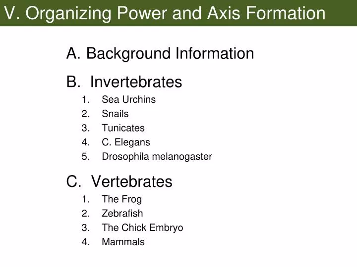
V. Organizing Power and Axis Formation. Background Information B. Invertebrates Sea Urchins Snails Tunicates C. Elegans Drosophila melanogaster C. Vertebrates The Frog Zebrafish The Chick Embryo Mammals. Part of these processes is the determination of axes in the organism

E N D
V. Organizing Power and Axis Formation • Background Information B. Invertebrates • Sea Urchins • Snails • Tunicates • C. Elegans • Drosophila melanogaster C. Vertebrates • The Frog • Zebrafish • The Chick Embryo • Mammals
Part of these processes is the determination of axes in the organism • The first few cleavages may produce little or no directionality to the embryo • It starts at varying stages in various animals and can result from different mechanisms
Figure 5.8 Fate map and cell lineage of the sea urchin Strongylocentrotus purpuratus
Step 1: Specification of Micromeres Two Big Changes: Specified to become skeletogenic mesenchyme Specified to become “Organizer” for other cells egg disheveled expression blocks B-catenin degradation
-catenin’s job NML All endo and meso ALL All ecto NONE
Step 2: “Organizing Power” • Secrete Wnt-8 into autocrine loop • Wnt-8Blimp-1B-cateninWnt-8 • Paracrine “early signal” induces macromeres and vegetal cells to differentiate to vegetal endoderm • Unknown signal as of yet • Delta-Notch juxtacrine signal induces non-skeletogenic mesenchyme • Wnt-8 makes a come-back to induce invagination
Axis Determination • Anterior-Posterior: Cytoplasmic determinants in the egg cytosol, such as disheveled and B-catenin • Left-Right: Nodal expression (TGF-B family member) • Dorsal-Ventral: unclear
Spiral cleavage in molluscs The spirally cleaving mollusks have a strong autonomous specification from cytoplasmic determinants in egg.
Step 1: Polar lobe formation The polar lobe is a cytoplasm outpouching from the egg prior to cleavage It isolates critical determinants into only one of the first cell pair. TF’s associated with the lobe turn CD into “The Organizer”
Figure 5.27 Association of decapentaplegic (dpp) mRNA with specific centrosomes of Ilyanassa Decapentaplegic is TGF-B family member used to induce specific cell fates secreted by the Organizer The Organizer induces mesodermal and endodermal fates in cells that would otherwise remain ectodermal
MAP kinase activity activated by D-quadrant snail blastomeres
Figure 5.30 MAP kinase activity activated by D-quadrant snail blastomeres (Part 2) Normal MAPK Blocked
Axis Determination • Anterior-Posterior: Cytoplasmic determinants in the lobe • Left-Right: Nodal expression (TGF-B family member) • Dorsal-Ventral: Cytoplasmic determinants in the lobe
Bilateral, Holoblastic Cleavage of the Tunicate The 8-cell embryo is already autonomously specified for cell fates
Figure 5.35 Cytoplasmic rearrangement in the fertilized egg of Styela partita Fertilization rearranges cytoplasmic determinants 1. Animal pole cytosol determines ectoderm 2. B-catenin presence determines endoderm (like urchins) 3. Macho-1 in yellow crescent determines muscle cells
Figure 5.38 Antibody staining of -catenin protein shows its involvement with endoderm formation Wherever B-catenin shows up, endoderm is formed
Figure 5.37 Autonomous specification by a morphogenetic factor Where Macho-1 shows up tail muscle will form Zinc-finger TF for muscle actin, myosin, TBX-6 Also TF for Snail TF which blocks notochord induction
Conditional Specification also plays a role Integrates with the autonomous specification patterns
Axis Formation accomplished prior to cleavage! Fertilization rearranges cytoplasmic determinants determines dorsal-ventral determines anterior-posterior Left-right: unclear but nodal shows it later
Rotational, Holoblastic Cleavage in the nematode Caenorhabditis elegans hermaphrodite
Figure 5.42 The nematode Caenorhabditis elegans (Part 2) Both autonomous and conditional specification at work early on. If cells are separated: P1 will develop autonomously Stem cell divisions are meridional Founder cell divisions are equatorial AB requires input from P lineage
Autonomous specification in P1 • SKN-1, PAL-1 and PIE-1 TFs from egg • as P1 divides these determine daughter fates • P lineage becomes “Organizer” • Conditional specification in AB • P2 secretes Wnt family member MOM-1 to induce endodermal specification in AB lineage • P2 use Delta-Notch signals to induce ectodermal fates in AB lineage
Axis Determination in C. elegans Anterior-Posterior axis is determined by egg shape Which end is posterior is determined by sperm (the closest end is back) Sperm CYK-4 activates egg rho, actin rearrangement causes assymetric first cleavage division
AB division leads to both dorsal ventral and left-right axes Assymetrical division of AB-MS forces AB dorsal and MS ventral Delta-notch recognition between daughters of AB and MS gives left-right
Cytoskeletal rearrangement also pushes P-granules into the germ line
The cells of the blastula have specified fates in Xenopus..... Gastrulation changes all of that, .....afterwards all cell fates are determined!
Development of “Organizing Power” at the dorsal blastopore lip The bottle cells get the ball rolling but the real power is conferred on the first cells through the blastopore.
The dorsal mesoderm keeps the power to determine other cell’s fates throughout gastrulation: “Spemann’s Organizer” This ability to determine cell fates is called... “Primary Embryonic Induction”
The dorsal lip cells first have to become competent to be “Organizer” Cortical rotation shifts disheveled, GBP, Wnt-11 to dorsal side of embryo The area of Dsh accumulation is seen as a gray crescent in some amphibian embryos
β-catenin starts out everywhere in the embryo but only survives GSK3 in the dorsal portion due to Dsh, GBP and Wnt-11
The dorsal vegetal cells of the Nieuwkoop Center turn on “Organizer” Wnt and Vg-1 (TGF-B family) induce pre-dorsal lip mesoderm FGF needed for all mesoderm
Figure 7.22 Summary of events hypothesized to bring about induction of the organizer in the dorsal mesoderm Nodal Vg-1
So, what can the “Organizer” do? • Initiate gastrulation • Become the notochord and other dorsal mesoderm • Dorsalize ventral mesoderm into paraxial mesoderm, somites, etc. • Dorsalize the ectoderm into the neural plate and neural tube
Figure 7.26 Localization of chordin mRNA The “Organizer” is induced prior to gastrulation Dorsal blastopore lip Blastopore Dorsal mesoderm Continues to organize events throughout its own differentiation
Interestingly, the primary mechanism is by means of inhibition....
Presumably, the Wnt, FGF and RA signals arise from endoderm and ectoderm Without the “Organizer” you get mainly skin and gut
Figure 7.31 Cerberus mRNA injected into a single D4 blastomere of a 32-cell Xenopus embryo induces head structures as well as a duplicated heart and liver Don’t underestimate the power of the “Organizer”!
Axis Formation • Dorsal-Ventral: sperm penetration and cortical rotation • Anterior-Posterior: migration direction of the dorsal mesoderm • Left-Right: nodal expression exclusively on left side of the lateral plate mesoderm
Nodal expression causes Pitx2 expression Nodal and Pitx2 on left Injected on both sides
Relationships between the frog and chick “Organizers” • The hypoblast = dorsal vegetal cells • Koller’s sickle = pre-dorsal lip mesoderm • Hensen’s node = dorsal blastopore lip and dorsal mesoderm • Primitive streak = blastopore
Formation of Hensen’s node from Koller’s sickle Wnt and FGF from the hypoblast induce Koller’s sickle epiblast
Figure 8.10 Induction of a new embryo by transplantation of Hensen’s node (Part 1)
Possible contribution of inhibition of BMP signaling Appears to be similar to the frog....
In the chick, the hypoblast plays a large role much like the frog endoderm
Figure 8.8 Specification of the chick anterior-posterior axis by gravity Anterior-Posterior axis parallels the rotation inside the shell
Rostral-Caudal (Anterior-Posterior) axis extension in chick embryos The combination of positional specification, complex signaling and TF (Hox, etc.) expression is thought to cause axis.
Left-right asymmetry in the chick embryo This is farther along Nodal and Pitx2 again are implicated