Download
1 / 43
440 likes | 775 Views
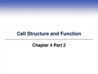
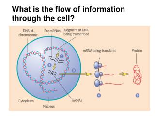
What is the flow of information through the cell?. Double helix - antiparallel polymers. Major groove Minor groove. 5’ 3’. A. T. G. C. Purine Pyrimidine. 06_12_asymmetrical.jpg. Transcription : dsDNA template Nucleotides (ACGU) make ssRNA Need to separate strands.

E N D
Double helix - antiparallel polymers Major groove Minor groove
5’ 3’
A T G C Purine Pyrimidine
Transcription: • dsDNA template • Nucleotides (ACGU) make ssRNA • Need to separate strands. • Nucleotides added to free 3’ OH (5’3’)
Classes of RNA… mRNA rRNA tRNA …also snRNA and microRNA
Prokaryotes untranslated regions 5’ UTR 3’ UTR promoter (not transcribed) coding sequence DNA mRNA RNA Polymerase Ribosome polypeptide
Eukaryotes untranslated regions 5’ URT 3’ UTR promoter (not transcribed) coding sequence DNA pre mRNA mRNA RNA Polymerase Ribosome polypeptide
Transcription start = +1 Consensus sequence = –35; TTGACA, recognized by Pribnow box = -10, TATAAT; determines +1 Terminator sequence: where polymerase stops Bacterial Promoter Elements
Initiation of eukaryotic transcription by RNA Pol II (mRNA) TF = transcription factor (compare with prokaryotic sigma factor)
Eukaryotic mRNA: • 5’ cap, • 5’ UTR • coding region • 3’ UTR • 3’ poly-A tail
mRNA processing in Eukaryotes 5’ cap added Remove 3’ end Poly-A tail added Introns removed Exons joined
replication transcription translation DNA RNA Protein Gene cloning - making lots of copies... 1. Make a “library” of small pieces of DNA (2 types) 2. Find the one piece you want 3. Insert it into a “vector” 4. Grow it in a new organism (bacteria, euk. cells) Isolate DNA, fragment with RE Isolate mRNA, convert to cDNA with reverse transcriptase Genomic library cDNA library
Overview of gene expression in eukaryotes 07_37_Protein.produc.jpg
Two adapters link an amino acid to a codon 07_26_2_adaptors.jpg
Intiation of translation in Eukaryotes 07_32_initiation.jpg
Intiation of translation in Prokaryotes 07_33_mRNA.encode.jpg
Elongation of proteins 07_30_3_step_cycle.jpg 4 5
07_34_stop codon.jpg Termination of translation
Frameshift: Adding or removing 1 or 2 nucleotides results in changes the reading frame from that point on. Nonsense: Changing an amino acid codon to a stop codon results in truncated proteins Missense: Changing an amino acid codon to one encoding a different amino acid - effect depends on type of amino acid and where in the protein. Mutations…
Primary structure (1°) of a protein: Arabidopsis -glucosidase (single letter codes) MSSLHWFPNIFIVVVVFFSLRSSQVVLEEEESTVVGYGYVVRSVGVDSNRQVLTAKLDLIKPSSVYAPDIKSLNLHVSLETSERLRIRITDSSQQRWEIPETVIPRAGNHSPRRFSTEEDGGNSPENNFLADPSSDLVFTLHNTTPFGFSVSRRSSGDILFDTSPDSSDSNTYFIFKDQFLQLSSALPENRSNLYGIGEHTKRSFRLIPGETMTLWNADTGSENPDVNLYGSHPFYMDVRGSKGNEEAGTTHGVLLLNSNGMDVKYEGHRITYNVIGGVIDLYVFAGPSPEMVMNQYTELIGRPAPMPYWSFGFHQCRYGYKNVSDLEYVVDGYAKAGIPLEVMWTDIDYMDGYKDFTLDPVNFPEDKMQSFVDTLHKNGQKYVLILDPGIGVDSSYGTYNRGMEADVFIKRNGEPYLGEVWPGKVYFPDFLNPAAATFWSNEIKMFQEILPLDGLWIDMNELSNFITSPLSSGSSLDDPPYKINNSGDKRPINNKTVPATSIHFGNISEYDAHNLYGLLEAKATHQAVVDITGKRPFILSRSTFVSSGKYTAHWTGDNAAKWEDLAYSIPGILNFGLFGIPMVGADICGFSHDTTEELCRRWIQLGAFYPFARDHSSLGTARQELYLWDSVASSARKVLGLRMRLLPHLYTLMYEAHVSGNPIARPLFFSFPQDTKTYEIDSQFLIGKSIMVSPALKQGAVAVDAYFPAGNWFDLFNYSFAVGGDSGKHVRLDTPADHVNVHVREGSIVAMQGEALTTRDARKTPYQLLVVASRLENISGELFLDDGENLRMGAGGGNRDWTLVKFRCYVTGKSVVLRSEVVNPEYASKMKWSIGKVTFVGFENVENVKTYEVRTSERLRSPRISLIKTVSDNDDPRFLSVEVSKLSLLVGKKFEMRLRLT
Secondary structure (2°) -helix H-bonds between C=O and N-H of backbone. (No R-groups involved)
Secondary structure -sheet H-bonds between C=O and N-H of backbone. (No R-groups involved)
Tertiary structure - the entire polypeptide -helix -sheet loops and turns disulfide bridge ribonuclease
lactic (lactate) dehydrogenase immunoglobulin light chain 04_20_protein domains.jpg cytochrome b562
Domains - discrete modules within tertiary structure that fold independently and have a specific function. 4 domains of phospholipase C
Motif - a recurring substructure / barrel e.g. -amylase
Motif - a recurring substructure coiled coil e.g. myosin
How do proteins get to their folded state? unfolded native conformation
Proteins with different functions may have similar shape - members of a family with a common ancestor. 04_21_Serine proteases.jpg