Download
1 / 1
10 likes | 61 Views
Nicole J. Huber and Gordon V. Wolfe, Dept. Biological Sciences, California State Univ. Chico njhuber@sbcglobal.net; gwolfe2@csuchico.edu. Introduction. Discussion. Results.

E N D
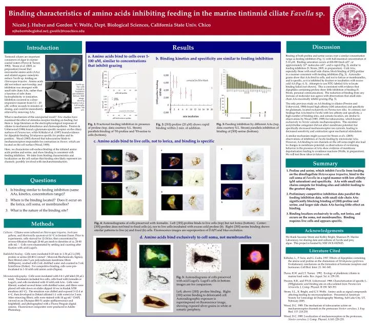
Nicole J. Huber and Gordon V. Wolfe, Dept. Biological Sciences, California State Univ. Chico njhuber@sbcglobal.net; gwolfe2@csuchico.edu Introduction Discussion Results Binding characteristics of amino acids inhibiting feeding in the marine tintinnid ciliate Favella sp. Binding of both proline and serine occurs over a similar concentration range as feeding inhibition (Fig. 1), with half-maximal concentration of 5-10 mM. Binding saturation occurs at 600-800 fmol cell-1, or approximately 1011 molecules cell-1, and is rapid (Fig. 2), similar to feeding inhibition (S. Strom, 2005, in preparation). Cold AAs, especially those with small side chains, block binding of [3H]-proline in a manner consistent with feeding inhibition (Fig. 3). Autoradio-grams show that AAs bind to cells, and not to loricas or membranelles, and is specific, as it is inhibited by fixation or incubation with excess cold AA (Figs. 4, 5). Attempts to use FITC-labeled AAs to locate binding failed (not shown). This is consistent with evidence that dipeptides containing proline show little inhibition of feeding (S. Strom, personal communication). The reduction of binding from any increase of molecular size agrees with observations that small side-chain AAs maximally inhibit grazing (Fig. 3). The only previous study on AA binding to ciliates (Preston and Usherwood, 1988) found high affinity (nM saturation) and specificity for glutamate, located exclusively on Paramecium cilia. In contrast, our findings that AAs bind to Favella with low affinity and specificity, a high number of binding sites, and somatic location, are similar to observations by Wood (1985, 1989) for tubocurarine, which bound exclusively to Stentor somatic mechanoreceptors. This nicotinic acetylcholine antagonist did not affect membrane potential, but specifically decreased mechanoreceptor ion currents, leading to decreased sensitivity and contraction upon mechanical stimulation. A similar mechanism might account for Strom et al’s. (2005) observations of inhibition of Favella feeding by micromolar AAs. However, AA binding to ion channels on the cell soma might also lead to changes in membrane potential, as observations of swimming behavior in the presence of AAs show evidence of membrane depolarization leading to avoidance reactions (Wolfe, in preparation). We will test these ideas in future work. Tintinnid ciliates are important consumers of algae in marine coastal waters (Pierce & Turner, 1992). Strom et al. (2005, in preparation) found that micromolar amino acids (AAs) and related organic osmolytes reduce Favella sp. feeding on Heterocapsa triquetra. Amino acids did not reduce survivorship, and inhibition was strongest with small side chain AAs, rather than a function of side chain hydrophobicity or ionization. Inhibition occurred in a dose-responsive manner from 0.2 – 20 mM, within seconds to minutes of dosing, and could be immediately relieved by removing AAs. b. Binding kinetics and specificity are similar to feeding inhibition a. Amino acids bind to cells over 1-100 uM, similar to concentrations that inhibit grazing Favella sp. Top: cells viewed in brightfield and darkfield. Bottom: SEM image, showing hyaline lorica. Cell lengths approx. 180 mm. What is mechanism of this unexpected result? Few studies have examined the effect of stimulus-receptor binding on feeding, but there is large literature on the electrophysiological responses of ciliates to mechanical disturbance and chemorepellents. Preston & Usherwood (1988) found a glutamate-specific receptor on the ciliary surfaces of Paramecium, while Köhidai et al. (1997) found evidence for dipeptide binding Tetrahymena specific for proline and its position. Wood (1985) showed that tubocurarine binds to mechanoreceptors responsible for contraction in Stentor, which are located on the cell surface (Wood, 1989). Here, we characterize cell-surface binding of the tritiated amino acids proline and serine, and show binding is consistent with feeding inhibition. We infer from binding characteristics and localization on the cell surface that binding sites likely represent ion channels, possibly involved with mechanotransduction. Fig. 2: [3H]-proline (20 mM) shows rapid binding within 2 min. of addition. Fig. 3: Feeding inhibition by different AAs (top, data courtesy S.L. Strom) parallels inhibition of binding of [3H]-serine (bottom). Fig. 1: Fractional feeding inhibition in presence of proline (top, data courtesy S.L. Strom) parallels binding of 3H-proline and 3H-serine to cells (bottom). c. Amino acids bind to live cells, not to lorica, and binding is specific Summary • Proline and serine, which inhibit Favella from feeding on the dinoflagellate Heterocapsa triquetra, bind to the cell soma of Favella in a rapid manner with low affinity (mM saturation) and specificity. AAs with small side chains compete for binding sites and inhibit feeding to the greatest degree. • Preliminary competitive inhibition data parallel the feeding inhibition data, with small side chain AAs significantly blocking binding of [3H]-proline and serine, and larger side chain AAs having little effect on binding. • Binding localizes exclusively to cells, not lorica, and occurs on the soma, not membranelles. Binding requires live cells and appears specific. Questions • Is binding similar to feeding inhibition (same AAs, kinetics, concentration range)? • Where is the binding located? Does it occur on the lorica, cell soma, or membranelles? • What is the nature of the binding site? Methods Fig. 4: Autoradiograms of cells preserved with formalin. Left: [3H]-proline binds to live cells (top) but not lorica (bottom). Center: [3H]-proline does not bind to fixed cells (a), nor to live cells incubated with excess cold proline (b). Right: [3H]-serine binding shows similar patterns to live (a) and fixed (b) cells. Fluorescence images are superposition of DAPI and blue excitation. Acknowledgements • Cultures - Ciliates were cultured on Heterocapsa triquetra, Isochrysis galbana, and Mantoniella squamata at 16 oC in Instant Ocean. Prior to experiments, cells starved for 12-24 hrs, then concentrated by reverse-filtration through 20-40 mm mesh to densities of ca. 20-80 cells mL-1. Cells were enumerated by settling and counting after fixation with acid Lugol’s. • Radiolabel binding - Cells were incubated 0-20 min in 1-30 mCi L-[3H] proline or serine (20-80 Ci mmol-1, Moravek Biochemicals, Sigma), then filtered onto 5 mm polycarbonate membrane filters (Millepore), washed with 2 mL distilled water and counted in 5 mL ScintiVerse (Fisher). For competitive binding, cells were pre-incubated in 1-10 mM cold amino acids (Sigma). • Microautoradiography - Cells were incubated with 0.4-1 mM label (30 mCi total). Treatments included live cells, cells fixed with formalin or Lugol’s, and cells incubated with 10 mM cold AAs. Cells were filtered, washed several times with distilled water, and filters were placed cell-side down on slides dipped 10 sec in Kodak NTB emulsion (43 oC). The emulsion was chilled and exposed 5-12 d at 4 oC, then developed in Dektol (diluted 1:1 with water) for 2 min. After removing filters, cells were stained with 10 mg mL-1 DAPI, viewed on an Olympus BH-51 under epifluorescence and brightfield, and photographed with a Pixera Penguin digital camera. Fluorescence composites were produced in Adobe Photoshop. d. Amino acids bind exclusively to cell soma, not membranelles We thank Suzanne Strom and Kelley Bright, Shannon Pt. Marine Laboratory, for sharing data and cultures of Favella and prey algae. This project is funded by NSF OCE-0325025. Literature Cited Köhidai, L., P. Soós, and G. Csaba. 1997. Effects of dipeptides containing the amino acid proline on the chemotaxis of Tetrahymena pyriformis. Evolutionary conclusions on the formation of hormone receptors and hormones. Cell Biol. Inter. 21: 341-345. Pierce, R.W. and J.T. Turner. 1992. Ecology of planktonic ciliates in marine food webs. Rev. Aquat. Sci. 6: 139-181. Preston, R.R., and P.N.R. Usherwood. 1988. Characterization of specific L-[3H]glutamic acid binding site on cilia isolated from Paramecium tetraurelia. J. Comp. Physiol. B 158: 345-351. Strom, S.L., K. Bright, and G.V. Wolfe. Amino acids as signal compounds affecting feeding by microzooplankton. Presented at American Society for Limnology & Oceanography Meeting, Salt Lake City, UT, February 2005. Wood, D.C. 1985. The mechanism of tubocurarine action on mechanoreceptor channels in the protozoan Stentor coeruleus. J. Exp. Biol. 117: 215-235. Wood, D.C. 1989. Localization of mechanoreceptors in the protozoan, Stentor coeruleus. J. Comp. Physiol. A 165: 229-235. Fig. 5: Autoradiograms of cells preserved with acid Lugol’s. Lugol’s cells in bottom images are for comparison. Left, above: [3H]- proline binding. Right: [3H]-serine binding to deloricated cell. Autoradiographic exposure is superimposed on fluorescence image, showing exposed silver grains in white at somatic periphery.