Download
1 / 12
120 likes | 301 Views
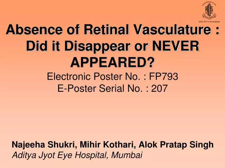
Absence of Retinal Vasculature : Did it Disappear or NEVER APPEARED? Electronic Poster No. : FP793 E-Poster Serial No. : 207. Najeeha Shukri, Mihir Kothari, Alok Pratap Singh Aditya Jyot Eye Hospital, Mumbai. Disclaimer. No financial interest or assistance has been received for this article.

E N D
Absence of Retinal Vasculature :Did it Disappear or NEVER APPEARED?Electronic Poster No. : FP793E-Poster Serial No. : 207 Najeeha Shukri, Mihir Kothari, Alok Pratap Singh Aditya Jyot Eye Hospital, Mumbai
Disclaimer • No financial interest or assistance has been received for this article.
Aim To report and discuss the Patho-embryology of an extremely rare case with Total absence of Retinal circulation in a child with Bilateral Optic Nerve Hypoplasia.
Introduction • Vasculogenesis is the formation of new blood vessels through de novo formation of new endothelial cells. • Angiogenesis is formation of vessel by budding & sprouting from already existing vessels. • Vasculogenesis of the retina begins around 15 weeks of gestation. Angiogenesisstarts at 21 weeks and is completed by term. Interruption during vessel development may cause - • PHPV • ROP • FEVR • Hypoplasia of optic nerve (rare case - described here). Here we describe a rare case of total absence of retinal vasculature.
Material & Methods • 3 month old child presented with leucokoria in the right eye. • 2nd child, normal full term delivery. • Non consanguinous marriage • No significant perinatal history. • Pedigree analysis shows - M F 32yrs 27yrs M M 3 yrs 3 months leucokoria
Examination revealedRight eye– Absent menace reflex, no nystagmus, myopia, hypoplastic optic nerve & retinal detachment. RE Microcornea (7.5mm) Posterior synechiae with Iris vascularisation Leukocoria Left eye – Absent menace reflex, no nystagmus, microcornea (7.5mm), chorioretinal coloboma, empty/pale optic disc, thready optic nerve (CT) LE LE Tesselated fundus Retinochoroidal coloboma Choroidal vasculature present Hypoplastic & empty optic disc. Absent retinal vessels.
BScan RE – Hyper reflective membrane attached to the disc s/o Retinal Detachment with SRF with no e/o calcification. CT Brain - Axial – Normal Septum Pellucidum, No associated intracranial midline defects / abnormality. CT - Axial section – Bilateral thready optic nerves.
DISCUSSION Normal development of hyaloid vasculature This case denotes an abnormality either in vasculogenesis, angiogenesis or related apoptosis which causes regression of fetal vasculature. Cross section 4-5wks 5wk Optic stalk Hyaloid artery 6-7wks Tunica Vasculosa lentis 6-7wk Hyaloid vasculature starts to regress by 2nd month & disappears completely by 8 month
DISCUSSION Normal development of retinal vasculature 3-4 mnth – primitive vascular mesenchyme cells appears near optic nerve 4-5 month - Solid endothelial cords sprout – differentiation and migration process. Angiogenesis starts Filopodial processess of endothelial cells seen 6th month faint lumen develops. 7- 8th month – vessels migrate outward 6-7months 8-9months • Stimulus for angiogenesis – • local hypoxia of neurons – VEGF produced by astrocytes and microglia • IGF- 1 causes proliferation of endothelial cells in hypoxic retinae. • PDGF
DISCUSSION • Regression of fetal vasculature is a known fact eg. Patent ductus arteriosus, renal vessels etc. Regression of retinal vasculature is mainly caused by apoptosis of the endothelial cells. Any untoward event during development can cause the following sequelae - • PHPV/PFV • FEVR – Premature arrest of development of peripheral retinal vasculature • Retinopathy of Prematurity • Absence / Incomplete vasculature • Few others • Mittendorfs dot • Bergmisters papillae • Vitreous cysts Mittendorfs dot ROP FEVR
Vasculature never appeared Complete absence of origin of retinal vessels. No persistance of remnants of retinal vessels at the origin i.e. on optic disc or glial cell proliferation / fibrous tissue. Appeared & later disappeared Presence of Normal transparent retina. Failure of mesoderm (hyaloid) invasion through the choroidal fissure primarily, retinal blood perfusion possibly through choroid. Complete vasculogenesis with later impedence of vasculature at the level of distal optic nerve as evidenced by hypoplastic nerves. CONCLUSION With this scenario we come to two conclusions : We feel the necessity of Retinal doppler, Fundus fluorescein angiography for detection of ghost vessels. MRI imaging for pituitary abnormalities & blood tests for endocrinal deficiencies are required. Further research is needed for knowing the exact reason for regression of vasculature / early apoptosis in this case.