Download

1 / 30
310 likes | 483 Views
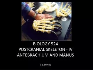
Antebrachium Flexors. Bony, soft landmarks . 1. humerus a. lateral epicondyle - radial side b. medial epicondyle - ulnar side 2. radius - head, neck, tuberosity, styloid process (more distal than ulnar's styloid)

E N D
Bony, soft landmarks • 1. humerus • a. lateral epicondyle - radial side • b. medial epicondyle - ulnar side • 2. radius - head, neck, tuberosity, styloid process (more distal than ulnar's styloid) • interosseous border (medial edge, attachment of interosseous mb); distal tubercle • 3. ulna - olecranon process, coronoid process & tuberosity, styloid process, trochlear notch interosseous border (lateral edge); head (distal end of ulna) • 4. flexor retinaculum: thick section of deep fascia at wrist -holds forearm tendons in place
Vessels • Brachial artery: extension/continuation of axillary artery (barely reaches into forearm) • Radial artery: lateral, anterior to elbow; partially overlapped by brachioradialis muscle • a. radial recurrent (around elbow, anastomoses with branch from deep brachial artery) • b. superficial palmar branch to form the superficial palmar arch (anastomoses with ulnar branch) • c. palmar carpal branch arch (joins with palmar carpal branch from ulnar artery to form the palmar carpal arch) • d. dorsal carpal branch (joins with dorsal carpal branch from ulnar artery to form dorsal arch, more later)
Ulnar artery • ulnar artery: medial, anterior to elbow, deep to pronator teres muscle • a. anterior ulnar recurrent anastomoses with inferior ulnar collateral artery • b. posterior ulnar recurrent anastomoses with superior ulnar collaterals around elbow • c. common interosseous artery. - in cubital fossa (branch into anterior, posterior branches) • d. palmar carpal branch arch (anastomoses with radial branch, see above) • e. dorsal carpal branch arch • f. superficial branch to form the superficial palmar arch • g. deep palmar branch arch (anastomoses with radial artery)
Forearm Nerves • Median & Ulnar - both are on the anterior side, supply essentially all the flexors with few exceptions • 1. Median - on anterior side • a. articular branches: to elbow • b. muscular branches: to all flexors (except flexor carpi ulnaris & medial part of digitorum profundus by ulnar nerve) • c. anterior interosseous nerve: to flexor digitorum profundus (lateral 1/2) pollicis longus & pronator quadratus • d. palmar cutaneous branch: - to skin of lateral palm
Ulnar • a. articular branches: to elbow • b. muscular branches: to flexor carpi ulnaris & medial part of digitorum profundus • c. palmar cutaneous branch: - to skin of posterior, medial part of hand
Radial Nerve • Radial - to posterior (supplies only extensors + brachioradialis, its one and only flexor) • a. superficial branch = continuation of radial- to skin on lateral dorsal wrist, hand, thumb, lateral 1+1/2 digits (index, middle) • b. deep branch - is larger, exclusively muscular & articular - to extensor muscles; supinator • c. posterior interosseous - a branch of the deep radial - to deep extensors
Muscles • 3 functional groups: • flex wrist at hand = flexor carpi radialis; flexor carpi ulnaris; palmaris longus • flex fingers = flexor digitorum superficialis; flexor digitorum profundus; flexor pollicis longus • pronate hand = pronator teres; pronator quadratus
layers • Superficial layer • pronator teres; flexor carpi radialis; flexor carpi ulnaris; palmaris longus • share a common tendon from humerus (medial epicondyle) • & deep fascia that covers proximal parts of the muscles • then separate from each other as they extend distal to the humerus • 2. intermediate layer = flexor digitorum superficialis • 3. deep layer = flexor digitorum profundus; flexor pollicis longus; pronator quadratus
PRONATOR TERES • ORIGIN • Origin of humeral head: Immediately above medial epicondyle of humerus, common flexor tendon, and deep antebrachial fascia • Origin of ulnar head: medial side of coronoid process of ulna • INSERTIONmedial of lateral surface of radius • ACTIONpronates forearm • NERVEmedian nerve – C6, 7
FLEXOR CARPI RADIALIS • ORIGINcommon flexor tendon from medial epicondyle of humerus, and deep antebrachial fascia • INSERTIONbase of second metacarpal bone and a slip to base of third metacarpal bone • ACTIONflexes, abducts the wrist and may assist in pronation of the forearm and flexion of the elbow • NERVEmedian nerve - C6, C7, C8
PALMARIS LONGUS • ORIGINcommon flexor tendon from medial epicondyle of humerus, and deep antebrachial fascia • INSERTIONflexor retinaculum, and palmar aponeurosis. • ACTIONtenses the palmar fascia, flexes the wrist, and may assist in flexion of the elbow • NERVEmedian nerve – C(6), C7, C8, T1
PALMARIS BREVIS • ORIGINulnar border of palmar aponeurosis and palmar surface of flexor retinaculum • INSERTIONSkin on ulnar border of hand • ACTIONCorrugates the skin on ulnar side of hand • NERVEulnar, C7, 8, T1
FLEXOR CARPI ULNARIS • ORIGIN • humeral head: common flexor tendon from medial epicondyle of humerus • ulnar head: by aponeurosis from the medial margin of olecranon, proximal two thirds of posterior border of ulna, and from the deep antebrachial fascia • INSERTIONpisiform bone and by ligaments, to hamate and fifth metacarpal bones • ACTIONflexes and adducts the wrist and may assist in flexion of the elbow • NERVEulnar nerve - C7, C8, T1
FLEXOR DIGITORUM SUPERFICIALIS • ORIGIN • Origin from humeral head: common flexor tendon from medial epicondyle of humrus, ulnar collateral ligament of elbow joint, and deep antebrachial fascia • Origin from ulnar head: medial side of coronoid process • Origin from radial head: obique line of radius • INSERTIONby four tendons into sides of middle phalanges of second through fifth digits • ACTIONflexes the proximal interphalangeal joints of second through fifth digits, assists in flexion of the metacarpophalangeal joints and in flexion of the wrist • NERVEmedian nerve - C7, C8, T1
FLEXOR DIGITORUM PROFUNDUS • ORIGINAnterior and medial surfaces of proximal three fourths of ulna, interosseus membrane, and deep antebrachial fascia • INSERTIONby four tendons into bases of distal phalanges, anterior surface • ACTIONflexes distal interphalangeal joints of index, middle, ring, and little fingers, and assists in flexion of proximal interphalangeal and metacarpophalangeal joints, may assist in flexion of the wrist • NERVEProfundus I, II median nerve C7, C8, T1Profundus III, IV: ulnar nerve C7, C8, T1
FLEXOR POLLICIS LONGUS • ORIGINAnterior surface of body of radius below tuberosity, interosseus membrane, medial border of coronoid process of ulna, and/or medial epicondyle of humrus • INSERTIONBase of distal phalanx of thumb, palmar surface • ACTIONflexes the interphalangeal joint of the thumb, assists in flexion of the metacarpophalangeal and carpometacarpal joints, and may assist in flexion of the wrist • NERVEmedian nerve, C(6),C7, C8, T1
PRONATOR QUADRATUS • ORIGINMedial side, anterior surface of distal one fourth of ulna • INSERTIONLateral side, anterior surface of distal one fourth of radius • ACTIONpronate the forearm • NERVEmedian nerve, C7, C8, T1