Download
1 / 64
640 likes | 645 Views
CUTANEOUS INFECTIONS AND INFESTATIONS. DR. MOHAMMED ALSHAHWAN MD. OBJECTIVES 1. General understanding of the causative organisms of common skin infection(CSI). 2. Focus on CSI clinical presentation. 3. Overview of the basic investigations done and general knowledge of first line therapy.

E N D
CUTANEOUS INFECTIONS AND INFESTATIONS DR. MOHAMMED ALSHAHWAN MD
OBJECTIVES 1. General understanding of the causative organisms of common skin infection(CSI). 2. Focus on CSI clinical presentation. 3. Overview of the basic investigations done and general knowledge of first line therapy.
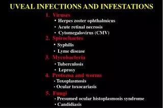
BACTERIA (impetigo , erysipelas &cellulits ) VIRUS (wart ,herpes simplex & herpes zoster) FUNGUS (Tinea , candidasis) PARASITE (Lieshmaniasis ,scabies & pediculosis)
BACTERIAL I. Impetigo Superficial non-follicular infection due to staphylococcus and streptococcus Children not sick pustule (honey-colored crust ) Face and Acral areas Primary or secondary
II. Erysipelas deep cutaneous infection (Dermal) due to streptococcus after penetrating trauma ( CHRONIC LYMPHEDEMA) sick Face and Acral areas Unilateral sharply demarcated edematous red plaque
III. Cellulitis deep cutaneous infection (up to SC FAT) due to streptococcus after penetrating trauma ( CHRONIC LYMPHEDEMA) sick Face and Acral areas Unilateral Diffuse (NOT well demarcated) edematous red plaque Blood Culture in immuocompramized pts.
VIRAL INFECTION WART Human papilloma virus (HPV) Direct contact Asymptomatic transmition Delay in presentation Oncogenic potential (HPV 16 and 18) High recurrence rate
CUTANOUS ( HPV 1 and 3 ) common wart flat wart planter wart GENITAL (HPV 6 and 11) classic condyloma acuminata
GENITAL WART *STD *Oncogenic HPVs ( Cervical cancer) *Usually more persistent and difficult to treat .
TREATMENT * Tissue destructive modalities Keratolytic (salicylic acid and podophyllin) Cryotherapy ( Liquid nitrogen) Electrotherapy CO2 laser * Immunotherapy
HERPES SIMPLEX Human Herpes virus I and II Direct contact Asymptomatic transmition Latency High recurrence rate
CUTANEOUS ( HSV I ) orolibialis Initial Herpatic whitlow Recurrence herpes ophtalmicus GENITAL ( HSV II ) Initial Recurrence
Incubation period : 7- 10 days. After 24-48 hours of burning and tingling sensation the patient develop grouped vesicles on erythematous base which ulcerate within 24 hours. The whole illness is around 7-10 days.
Tzank smear Direct fluorescent antibody test Viral culture Blood serology
VARICELLAE ZOSTER VIRUS (VZV) RESPIRATORY DROPLETS CHICKENPOX ( Children) HERPES ZOSTER (Adult) is due to reactivation of VZV which was dorminant in nerve root ganglion
CHICKENPOX Incubation period : 2 weeks Prodrom of respiratory coryza followed by disseminated red macules with central vesicles. The whole illness : 3 weeks The patient contagious 5 days before and 5 days after skin eruption
HERPES ZOSTER After 24-48 hours of burning and tingling sensation the patient develop grouped vesicles on erythematous base which ulcerate within 24 hours. The whole illness is around 7-10 days. Post-herpetic neuralgia (PHN) which usually persist for around 4 weeks.
It is almost always DERMATOMAL SPINAL (Thoracic ) CRANIAL ( Trigeminal) SERIOUS involvement 1.Ophthalmic division of trigeminal nerve. 2. Geniculate ganglia (Ramsey-hunt syndrome) 3.Sacral ganglia.
Treatment HERPES SIMPLEX Acyclovir 200 mg five time a day for a week HERPES ZOSTER Acyclovir 800 mg five time a day for a week
FUNGAL DERMATOPHYTE Tinea Pedis (most common) 1.Erosive interdigitalis 2. Hyperkeratotic type(T. rubrum) 3. Inflammatory type(T.mentagrophyte)
Tinea corporis / Tinea cruris 1.Hyperkeratotic type (T. rubrum) well-demarcated annular red hyperkeratotic plaque with central clearing (Ring worm) 2.Inflammatory type (T.mentagrophyte) well-demarcated edematous red plaque with superimposed pustules
Tinea Capitis 1.Hyperkeratotic (black dot) usually due to T. tonsurans 2. Inflammatory (Kerion) usually due to M. canis complex 3. Favus * Due to T. schoenleinii * it characterized by the presence of Scutulae .
YEAST Candidosis Due to candida albicans It is a commensal flora of the gut which become pathogenic when the immune status of the person changed
physiological (old age , neonate and pregnancy) pathological ( DM, HIV and organ transplant) Itrogenic (long course of Antibiotics)
MUCOSAL 1. Oral oral thrush angular chilitis 2. Genital valvuvaginitis
CUTANEOUS it favor wet areas Candidal intertrigo ( Napkin rash) peripherally spreading glazed red patch with scaly border and satellite pustules Candidal paronychia
pityriasis versicolor Due to Malassezia furfur Asypmtomatic Well-demarcated brown patches with branny over the trunk and upper extremities
1. Scraping,Clipping and Hair blucking KOH/microscopy Culture 2. Skin biopsy Histopathology with PAS stain Culture
Topical Antifungal Nystatin preparation (oral thrush) Imidazoles e.g. cotrimazole and miconazole Systemic Antifungal Imidazoles e.g. Itraconazole and fluconazole Allylamine e.g. Terbinafine Griseovulvin
PROTOZOA Lieshmaniasis Protozoa called Lieshmania Sand fly (premastigote) Macrophage (Amastigote) Lieshman-Donovan bodies
Localized Cutaneous Well-demarcated ulcerated nodule over the exposed areas after a trip to an endemic area ( H/o of insect bite) Disseminated Cutaneous Mucocutaneous Visceral