Download
1 / 94
950 likes | 1.8k Views
Review chest X-ray. By Elizabeth Kelley Buzbee AAS, RRT-NPS, RCP Lone Star College: Kingwood Respiratory Care Program. 1. In the chest x-ray of a person with pneumocystis carinii pneumonia (PCP) one would most likely see______ because this diseases progresses rapidly to ARDS.

E N D
Review chest X-ray By Elizabeth Kelley Buzbee AAS, RRT-NPS, RCP Lone Star College: Kingwood Respiratory Care Program
1. In the chest x-ray of a person with pneumocystiscarinii pneumonia (PCP) one would most likely see______ because this diseases progresses rapidly to ARDS. a. lobar consolidation b. pneumomatoceles c. Kerley B lines in the bases d. left-sided effusions e. diffuse tiny opacities & air bronchograms
answer e. diffuse tiny opacities & air bronchograms because this is what we see in an alveolar problem. We would see this in any diffuse pneumonia or in ARDS or IRDS.
2. In the chest X-ray of a person with staphylococcal pneumonia one would most likely see : a. lobar consolidation b. pneumomatoceles c. Kerley B lines in the bases d. left-sided effusions e. diffuse tiny opacities and air bronchograms
answer b. Pneumomatoceles a pneumomatocele is a air-filled cavity that shows up in 24 hours or less. They will be seen in staph pneumonia and in aspiration of hydrocarbons.
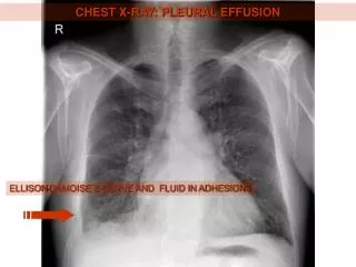
3. In the chest X-ray of the patient with left sided pleuritic pain and diminished LLL breath sounds, one might expect to see: a. LUL consolidation b. pneumomatoceles c. Kerley B lines d. left-sided effusions e. diffuse tiny opacities and air bronchograms
answer d. left-sided effusions Effusions will be seen as homogenous opacities that collect in the plural space in the dependent part of the thorax. One would have dullness to palpation and chest pain from the pluresy that would accompany the effusion
4. Kerley B lines are seen: I. when alveoli are full of fluid ii. when interstitial spaces are edematous iii. in tuberculosis iv. in pulmonary edema a. i and iv b. ii and iv c. iii only d. iii and iv
answer b. ii and iv Kerley B line are short, horizontal lines seen on the bases of the lung. The lines reflex thickened alveolar septal walls. They are seen in diffuse interstitial disorders such as interstitial pneumonia, pulmonary edema [interstitial and alveolar filling patterns]
5. One would see an air bronchogram at the level of the RML in an area of: a. consolidation b. air trapping c. atelectasis d. abscess
answer • Consolidation Consolidation is an alveolar filling pattern where the air is replaced by fluid. Fluid is white [opaque] on the X-ray Because the airway is black, we now see the airway against the white opacities
6. Persons with emphysema would most likely have the following on chest film: a. right-sided pleural effusion b. bullea/ bleb c. Kerley B lines d. abscess
answer • b. bullea/ bleb • Is an air pocket, seen in serious airtrapping. These will be seen in empysema and COPD.
7. Within a few hours of the incident, a baby who has aspirated a toy would most likely have distal to the obstruction: a. consolidation b. a cavitation c. atelectasis d. abscess e. a or c are both possible
answer e. a or c are both possible If the baby inhales a foreign object that completely cuts off gas flow to the lower airways, then there will be atelectasis If the object only causes a ball-valve obstruction, the there can be localized airtrapping below the object
8. If one sees homogenous opacities in the RML & narrowed intercostal spaces overlying this area, one is seeing: a. RML consolidation b. localized RML airtrapping c. RML atelectasis d. a RML mass
answer c. RML atelectasis One of the signs of atelectasis is a movement of adjacent objects into the place where the lung has collapsed. Ribs will be closer together and fissures may move toward the atelectasis While atelectasis is opaque like consolidation, there will be no air-bronchograms in atelectasis It is possible to have both atelectasis and consolidation in the same patient with many alveolar disorders
9. A thick walled opacity with an air/fluid interface is most likely: a. consolidation b. a mass c. atelectasis d. abscess e. b or c are possible
answer d. abscess An abscess is a thick-walled opacity that is filled with pus. It is caused by a necrotizing bacterial pneumonia. If the abscess ruptures into an airway, we might see the air/fluid interface inside the abcess
10. A tumor compressing the RUL bronchus could result in: a. RUL abscess b. RUL consolidation c. RUL atelectasis d. both a and b e. both b and c
answer e. both b and c Just like the F.O. a tumor compressing the airway can cause atelectasis & consolidation downstream If the tumor is smaller, it might cause a ball valve obstruction and result in a localized airtrapping and you might hear a wheeze over one spot—that will not respond to bronchodialators
11. When compared to viral pneumonia, bacterial pneumonias are more associated with: i. diffuse alveolar opacities with air bronchograms ii. localized alveolar opacities with air bronchogram iii. abscesses iv. cavitations a. i, iii b. ii, iii c. ii, iii and iv d. i only
answer c. ii, iii and iv Viral pneumonias tend to be diffuse, while bacterial pneumonias will be characterized by local problems such as abscesses, effusions or cavitations
12. Immediately after drainage of a small right-sided empyema by needle aspiration, a AP chest film is ordered. You see an area of hyperlucency without lung markings in the RUL. The heart shadow is almost completely to the left of the sternum. What has happened? a. the needle has successfully aspirated the fluid from the empyema b. the needle was too small, the fluid is too thick and the aspiration attempt was not successful. A chest tube must be inserted. c. the needle has punctured the lung and a tension pneumothorax has resulted
answer c. the needle has punctured the lung and a tension pneumothorax has resulted The heart has shifted, and the hyperlucency is air in the chest. This is a common hazard of thoracentesis. All procedures involving a needle and the chest must be followed by a chest x-ray to rule out pneumothorax
13. Immediately after the insertion of a flow directed pulmonary artery catheter, a chest film is ordered. You see a radiopaque line enter the right subclavian vein and you see that the tip of the catheter is in the right atrium. a. the catheter has migrated into the wedged position b. the catheter is not inserted far enough c. the catheter is in the proper position d. the catheter has transected the right subclavian vein
answer • c b. the catheter is not inserted far enough The RA is an excellent position for a central line but the pulmonary artery catheter should sit in the pulmonary artery
14. Immediately after a right-sided chest tube has been removed from the 3rd intercostal space in the anterior, a chest film is ordered. You see that there are lung markings from the hilar down to the pleura in the right apical area. This probably means that: a. the effusion has returned, the chest tube may need to be reinserted. b. the pneumothorax has returned, the chest tube may need to be reinserted. c. there in now a pulmonary infarction in this are d. the pneumothorax has resolved
answer d. the pneumothorax has resolved You want to see lung markings all the way to the plural. We know that the most common site for a chest tube to drain a pneumothorax is in the anterior upper chest
15. Signs of cardiogenic pulmonary edema include the following: i. Kerley B lines ii. cardiomegaly iii. increased opacities in the hilar area, in a butterfly pattern iv. segmental airtrapping a. i, ii, iii and iv b. ii, iii only c. i, ii only d. i, ii, iii
Answer d. i, ii, iii In all pulmonary edema we will see alveolar filling patterns and thickened alveolar septal walls, but if the heart is enlarged, it is cardiogenic pulmonary edema Another sign it is cardiogenic is the butterfly pattern [or bat wing] created by engorged pulmonary arteries
16. Air bronchograms are seen in cases of alveolar consolidation, because the opacity of the consolidation creates a contrast to the radiolucency of the airway as it lies over the area of consolidation. a. true b. false
Answer a. true Just like a black cat disappears in a dark room, we don’t normally see the black airways against black [air filled] alveoli if the alveoli are filled with fluid we now see the black airways against the opaque
Diffuse lesions of tiny opacities of less than 4 mm in diameter are seen in varicella pneumonia. This is also seen in: • diffuse pulmonary tuberculosis • multiple fat emboli c. ARDS d. no other lung disorder
Answer • diffuse pulmonary tuberculosis When one has dissiminated TB, there are tiny opacities that look like millet seeds. This is seen in chicken pox pneumonia also…very bad sign
18. It is not possible for one to have a combination of diffuse interstitial and alveolar filling patterns in the same patient who is diagnosed with non-cardiogenic pulmonary edema. a. true b. false
Answer • False • Because both interstitial and alveolar filling patterns are seen in problems with the alveoli, you can have both show up in the X-ray. These persons will have low compliant lungs and refractory hypoxemia
19. A person who has flattened diaphragms with wide intercostal spaces and bronchial thickening would most likely have: a. emphysema b. bilateral pneumothorax c. bilateral effusions d. a lobectomy e. pulmonary infarction
Answer a. emphysema the intercostal spaces are widened by airtrapping, the bronchial walls are thickened by secretions and the diaphragm has been pushed down by the airtrapping
20. The mediastinal structures tend to shift towards a : a. pneumothorax b. an area of airtrapping c. an area of atelectasis d. an area of consolidation e. c and d
Answer • c. an area of atelectasis • Structures move away from pneumothorax or from airtrapping • Nothing moves in consolidation
21. Mr. Reese had a LLL lobectomy. Several months after this surgery, one would not be surprised to find what abnormal findings on a PA chest film ? a. the LUL seems smaller or seems pulled to the right b. the LUL seems to be larger and it's inferior borders seem to bulge into the space where the LLL used to be. The heart seems to be more on the left than normal c. there is a hyper-lucency in the LLL and the left hemi-diaphragm is depressed d. there will be no changes
Answer b. the LUL seems to be larger and it's inferior borders seem to bulge into the space where the LLL used to be. The heart seems to be more on the left than normal Remember: objects move into a vacuum when the lung is removed, the other lobes move into the area—the fissures will be altered
22. When looking at a RUL pneumothorax, one would expect to see: i. hyperlucency without lung markings in the RUL ii. hyperlucency without lung marking in a column on the right side of the heart. iii. the superior aspect of the RML may be opaque iv. the 3rd-5th right intercostal spaces will be closer together than the same intercostal spaces on the left a. i, iii only b. ii, iii only c. i, iv only d. i, iii and iv
Answer • a. i, iii only • Pneumthorax always show up as hyperlucency • The air would push on the superior aspect of the RML so that it starts to collapse. It will become opaque
23. When the right hemi-diaphragm is paralyzed, one would see what derangement on the PA chest film during the inspiratory phase? a. the right hemi-diaphragm is 2 cm higher than the left b. the right hemi-diaphragm is 2 cm lower than the left c. the right hemi-diaphragm is 4 cm higher than the left d. the right hemi-diaphragm is 4 cm lower than the left
Answer c. the right hemi-diaphragm is 4 cm higher than the left • This is tricky. Remember: the normal position for the right hemi-diaphragm is to be 2 cm higher than the left. When the diaphragm is paralyzed, it sits in the resting position which is up so the right is higher than it should be.
24. A homogenous opacity located in the basal aspect of the LLL which causes the costophrenic angle to be blunted would most likely be a (an): a. LLL consolidation b. LLL atelectasis c. left sided effusion d. none of these
Answer • c. left-sided effusion • Blunting of the costophrenic angle is caused by fluid in the plural cavity. Fluid is opaque.
25. If you were to see an area in the LUL, which you would describe as a sharp and distinct round opacity you might be describing a/an: a. cavitation b. pneumatocele c. bullae or bleb d. abscess without an air/fluid interface e. an abscess with an air/fluid interface