Download
1 / 32
500 likes | 960 Views
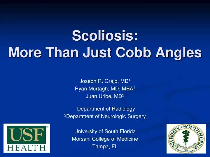
Scoliosis: More Than Just Cobb Angles. Joseph R. Grajo, MD 1 Ryan Murtagh, MD, MBA 1 Juan Uribe, MD 2 1 Department of Radiology 2 Department of Neurologic Surgery University of South Florida Morsani College of Medicine Tampa, FL. Disclosures. Joseph R. Grajo, MD: None

E N D
Scoliosis: More Than Just Cobb Angles Joseph R. Grajo, MD1 Ryan Murtagh, MD, MBA1 Juan Uribe, MD2 1Department of Radiology 2Department of Neurologic Surgery University of South Florida Morsani College of Medicine Tampa, FL
Disclosures Joseph R. Grajo, MD: None Ryan Murtagh, MD, MBA: None Juan Uribe, MD: Nuvasive® (consultant, research grants, speaker bureau, royalties) - no stocks
Goals and Objectives Introduce a 3-dimensional approach to scoliosis Discuss the various spinal and pelvic imaging parameters that play a significant role in patient outcomes after spinal surgery Discuss surgical approaches used to restore normal coronal and sagittal balance with representative pre- and post-operative cases
Target Audience General Radiologists Neuroradiologists
Scoliosis • Historically, surgeons have focused on coronal alignment (scoliosis) and surgery to correct coronal deformity • Recent findings show that sagittal alignment correlates highly with quality of life scores2 and that failure to correct sagittal deformity can lead to poor outcomes • We are also finding that the position of the pelvis plays a significant role in spinal alignment • As a result, there is a more global, 3-dimensional approach to spinal deformity with increased emphasis on correction of both spinal and pelvic deformities 2Glassman, et al, Spine (Phila Pa 1976) 30:682–688, 2005 1Dubousset “Cone of Economy” taken from Ames, et al, J Neurosurg: Spine / March 23, 2012
Spinal parameters – Pelvic parameters Spinal imaging parameters • Coronal spinal parameters • Scoliosis - measured by Cobb angles • Coronal plane decompensation • Sagittal spinal parameters • Thoracic Kyphosis (TK) • Lumbar Lordosis (LL) • Spinal Vertical Alignment (SVA)
Spinal parameters – Pelvic parameters Cobb angles 26 degrees of dextroscoliosis
Spinal parameters – Pelvic parameters Coronal Plane Decompensation Plumb line Central sacral vertical line • Measure by • Drawing plumb line from C7 midpoint • Then measure distance from plumb line to midpoint of S1 • In neutral, plumb line intersects midpoint of sacrum +/- 2 cm right or left Ames, et al, J Neurosurg: Spine / March 23, 2012
Spinal parameters – Pelvic parameters Coronal plane decompensation 4.6 cm
Spinal parameters – Pelvic parameters Thoracic Kyphosis • Normal is 30-40o in males 50-80 years old (increases with age) • Approx 40o in women of same age • Fon, et al, AJR 1980 Ames, et al, J Neurosurg: Spine / March 23, 2012
Spinal parameters – Pelvic parameters Lumbar Lordosis • Average in adults is 33.2 +/- 12.1 degrees • Increases with age • Lin, et al, J Formos Med Assoc, 1992 Ames, et al, J Neurosurg: Spine / March 23, 2012
Spinal parameters – Pelvic parameters Spinal Vertical Alignment 3Jackson and McManus. Spine 1994;19:1611–8 4Schwab, et al. Spine (Phila Pa1976) 35:2224–2231, 2010 6Malfari, et al, AJR 2010;194:S8–S22 • Measured as: • Distance between plumb line from mid point of inferior endplate of C7 and supero-posterior corner of S1 • In neutral SVA, plumb line from C7 intersects with supero-posterior corner of S1 • Mean in asymptomatic adults is 0.5 cm +/- 2.5 cm3 • Ideal SVA is less than +/- 5 cm4 • Increases with age (“anterior imbalance”) Ames, et al, J Neurosurg: Spine / March 23, 2012
Spinal parameters – Pelvic parameters Normal and abnormal SVA Normal/Neutral Positive SVA
Spinal parameters – Pelvic parameters Pelvic parameters • Parameters in adjacent zones of the spinopelvic axis are interdependent • Pelvic zone affects the lumbar zone (and vice-versa) • Lumbar zone affects the thoracic zone5 • Position of pelvis plays important role in upright sitting and standing postures • Failure to address abnormal pelvic alignment increases risk of spinal malalignment, decompensation and treatment failure • Pelvic parameters consist of • Pelvic Incidence (PI) • Pelvic Tilt (PT) • Sacral Slope (SS) • Pelvic Obliquity (PO) 5Berthonnaund, et al. J Spinal Disord Tech 2005;18:40–7.
Spinal parameters – Pelvic parameters Pelvic Incidence Ames, et al, J Neurosurg: Spine / March 23, 2012 • Angle between line from femoral head(s) to midpoint of sacrum and line perpendicular to superior endplate of sacrum • This is a morphologicparameter – descriptor of how much sacrum is angled in a person. Basically, it describes the shape of the sacrum that you were born with • Stays constant throughout life except for slight change in puberty • Important for its relationship to lumbar lordosis (LL) • Ideally LL = PI +/- 9 degrees3 • Can use a patient’s PI as pre-operative guide for amount of lordosis that should be introduced at surgery 3Schwab, et al. Spine (Phila Pa1976) 35:2224–2231, 2010
Spinal parameters – Pelvic parameters Pelvic Tilt 1Ames, et al, J Neurosurg: Spine / March 23, 2012 • Angle between a line from midpoint of femoral heads to center of superior endplate of sacrum and a vertical plumb line to midpoint of femoral heads • This is a positional parameter, meaning that unlike the PI, it can change • Compensatory mechanism – as spine tilts forward (i.e. as you lose lordosisand increase kyphosis), subject tries to maintain neutral alignment by pelvic retroversion4 • Increased work to retrovertpelvis and patients become uncomfortable leading to poor outcomes • Ideally PT is less than 20 degrees3 • Increased PT after surgery implies residual postoperative spinal deformity and negatively affects function and thus postoperative outcomes(highly correlated with outcomes)3 3Schwab, et al. Spine (Phila Pa1976) 35:2224–2231, 2010 4Jackon and McManus. Spine 1994;19:1611–8
Spinal parameters – Pelvic parameters Pelvic Tilt • Recall that parameters in adjacent zones of the spinopelvic axis are interdependent • Therefore As SVA increases, the result is a compensatory increase in PT • Study by Schwab, et al6 on patients compared changes in SVA vs pelvic tilt • Negative SVA (<0) – average PT = 10o • Neutral SVA (0-5 cm) – average PT = 16o • Positive SVA (> 5 cm) – average PT = 21o 6Schwab, et al, Spine 2006;31:E959–67.
Spinal parameters – Pelvic parameters Increasing PT to maintain neutral Low pelvic tilt but positive SVA Now has normal SVA but high PT, lots of energy to maintain Retroversion Increasing SVA increase in PT greater energy expenditure and greater disability Image from Ames, et al, J Neurosurg: Spine / March 23, 2012
Spinal parameters – Pelvic parameters Two examples of abnormal PT 35 degrees 31 degrees (note positive SVA) *this is good example showing that sometimes it is hard to align femoral heads. Attempt to use midpoint if not aligned.
Spinal parameters – Pelvic parameters What is the significance??? • Retroverted/compensated patients are not happy patients • Patients with positive sagittal balance fight to maintain neutral by • Pelvic retroversion • Flex hips and knees • More work to stay upright!! • Patients with sagittal balance + scoliosis had better outcomes when both fixed versus just scoliosis fixed7 • Another study showed best radiographic predictor of outcome was not scoliosis curve but sagittal balance8 7Schwab, et al, Spine 2008; 33:2243–2247 8Glassman, et al, Spine 2005; 30:682–688
Spinal parameters – Pelvic parameters Sacral Slope Ames, et al, J Neurosurg: Spine / March 23, 2012 • Measurement that is not as commonly used • Angle between line drawn along superior endplate of S1 and a horizontal reference line • PI = PT + SS • As PT increases, SS decreases (pelvic retroversion)
Spinal parameters – Pelvic parameters Summary of pelvic parameters by age 3Schwab, et al. Spine (Phila Pa1976) 35:2224–2231, 2010
Spinal parameters – Pelvic parameters Pelvic obliquity Important part of coronal correction strategy In similar fashion to the relationship between PT/retroversion in sagittal balance, PO is compensatory mechanism to improve coronal balance Presence of PO can suggest other problems like leg length discrepancy, and these should be addressed if present Ames, et al, J Neurosurg: Spine / March 23, 2012
Spinal parameters – Pelvic parameters Pelvic obliquity • Pelvic obliquity (tilt) to the right as compensatory mechanism to maintain neutral coronal balance
Spinal parameters + Pelvic parameters = The Big Picture How is positive balance/retroversion treated surgically? Flat back – before surgery Post op - increased lordosis with osteotomies and cages restores sagittal balance • With positive SVA, neutral sagittal balance can be restored through increase in lumbar lordosis • Lordosis can be increased by: • taking away height from the back (osteotomy, such as pedicle subtraction osteotomy) • adding height to the front (ALIF with introduction of cages to increase height) • Location can have variable results • More inferior – greater increase in LL with osteotomy or cage • More superior – requires larger osteotomy or cage for same lordosis Ames, et al, J Neurosurg: Spine / March 23, 2012
Before and After AFTER • Osteotomies increase LL from 28o to 60o • SVA of 3 cm • PT of 9o BEFORE Positive sagittal balance of 12 cm PT of 22o Ames, et al, J Neurosurg: Spine / March 23, 2012
Before and After Dextroscoliosis 55o Pelvic obliquity 7o Dextroscoliosis 19o Pelvic obliquity 5o
Before and After SVA 10.9 cm SVA 3.4 cm
Before and After Pelvic tilt 35 degrees Pelvic tilt 22 degrees
Bringing it all together • In summary, like it or not, we are in an age where radiology is fighting relevancy in many major areas • Imperative that radiologist develop larger role in surgical decision making process • A little extra effort on something as simple as scoliosis series can go a long way • As the radiologist, you should address the following in your report for scoliosis films • On the frontal view - where is the scoliosis and what are the Cobb angles of the primary and secondary curves? • How much coronal plane decompensation is there and is there any compensatory pelvic obliquity? • On the lateral view, what is the SVA and how much is the pelvic tilt? Conclude if there is compensation/pelvic retroversion (PT > 20o). • Is there a significant difference between the lumbar lordosis and the pelvic incidence? LL should be within 9o of the PI. If significant difference, can increase LL with osteotomy and/or cages.
Thanks! • Contact Information • Joseph R. Grajo, MD: jgrajo@health.usf.edu • Ryan Murtagh, MD: rmurtagh13@gmail.com • Juan Uribe, MD: juribe@health.usf.edu