Download
1 / 40
400 likes | 528 Views
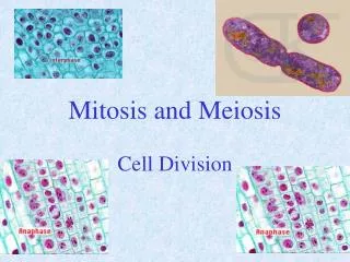
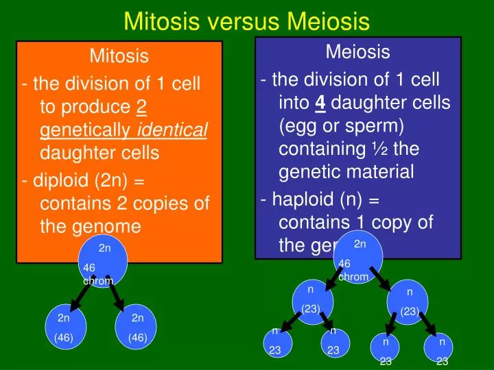
Mitosis - the division of 1 cell to produce 2 genetically identical daughter cells - diploid (2n) = contains 2 copies of the genome. Meiosis - the division of 1 cell into 4 daughter cells (egg or sperm) containing ½ the genetic material - haploid (n) = contains 1 copy of the genome.

E N D
Mitosis - the division of 1 cell to produce 2 genetically identicaldaughter cells - diploid (2n) = contains 2 copies of the genome Meiosis - the division of 1 cell into 4 daughter cells (egg or sperm) containing ½ the genetic material - haploid (n) = contains 1 copy of the genome Mitosis versus Meiosis 2n 46 chrom 2n 46 chrom n (23) n (23) 2n (46) 2n (46) n 23 n 23 n 23 n 23
Development of male and female gametes The Formation of sex cells during meiosis is referred to as gametogenesis Sperm and egg production are different: • In males 4 viable sperm are produced Spermatogenesis • In females 3 of the cells produce are known as polar bodies and do not survive. Only one egg is formed Oogenesis
Differences • The cyctoplasm of the female oocyte does not divide equally; one of the daughter cells called an ootid, receives most of the cyctoplasm while the other cells called polar bodies (“nurse cells”) die and are reabsorbed to provide nutrients • Sperms show equal division of cytoplasm; all 4 daughter cells become viable sperm
Spermatogenesis • Occurs in the seminiferous tubules where diploid spermatogonia, stem cells that are the precursors of sperm. • Spermatogonia • divide by mitosis to produce more spermatogonia or • differentiate into spermatocytes
Spermatogenesis • Meiosis of each spermatocyte produces 4 haploidspermatids. This process takes over three weeks to complete • Then the spermatids differentiate into sperm, losing most of their cytoplasm in the process.
Sketch Spermatogeneis showing meiotic divisions 46 • Spermatogonium 2. 1° Spermatocyte 3. 2° Spermatocyte 4. Spermatid 5. Sperm 1st Meiotic division 46 23 23 2nd Meiotic division 23 23 23 23
Oogenesis • Egg formation takes place in the ovaries. • the initial steps in egg production occur prior to birth. • Diploid stem cells called oogonia divide by mitosis to produce more oogonia and primary oocytes
Sketch Oogeneis showing meiotic divisions 46 • Oogonium 2. Oocyte • a) 2° Oocyte b) 1st polar body • a) Ootid b) polar bodies 5. Ovum 46 1st Meiotic division 23 23 2nd Meiotic division
By the time the fetus is 20 weeks old, the process reaches its peak and all the oocytes that she will ever possess (~4 million of them) have been formed. • By the time she is born, 1–2 million of these remain. Each has begun the first steps of the first meiotic division (meiosis I) and then stopped.
No further development occurs until years later when the female becomes sexually mature. • Then the primary oocytes recommence their development, usually one at a time and once a month. Unfertilized oocyte
The primary oocyte grows much larger and completes the meiosis I, forming a large secondary oocyte and a small polar body that receives little more than one set of chromosomes. Which chromosomes end up in the egg and which in the polar body is entirely a matter of chance.
In humans, 22 of the 23 are homologous; these are the autosomes • Thomas Hunt Morgan discovered that having 2 rod shaped (X chromosome) indicated female and 1 rod with 1 hooked shaped chromosome (Y chromosome) identified males • The X and Y are not homologous, they are the sex chromosomes
Meiosis and Variation • Unlike mitosis, meiosis does not produce identical cells • The cells produced only have half the number but the chromosomes therefore only ½ the genetic information • What chromosomes end up in what cell all depend upon how the chromosomes line up in Metaphase I
Meiosis and Variation • If the two blue chromosomes line up on the same side and the two red chromosomes line up on the same side • Then the daughter cells will have either the genetic information from the red or blue chromosome
Meiosis and Variation • If 1 red and 1 blue line up on the same side • The daughter cells will have genetic information from both red and blue chromosomes
MITOSIS MEIOSIS PARENT CELL(before chromosome replication) Site ofcrossing over MEIOSIS I PROPHASE I Tetrad formedby synapsis of homologous chromosomes PROPHASE Chromosomereplication Chromosomereplication Duplicatedchromosome(two sister chromatids) 2n = 4 Chromosomes align at the metaphase plate Tetradsalign at themetaphase plate METAPHASE I METAPHASE ANAPHASETELOPHASE Sister chromatidsseparate duringanaphase Homologouschromosomesseparateduringanaphase I;sisterchromatids remain together ANAPHASE I TELOPHASE I Haploidn = 2 Daughtercells of meiosis I No further chromosomal replication; sister chromatids separate during anaphase II MEIOSIS II 2n 2n Daughter cellsof mitosis n n n n Daughter cells of meiosis II
Abnormalitiesduring Meiosis The movement of the chromosomes in a dividing cell is so precise that only 1 in every 100,000 divisions will contain an error Non-disjunction = an error during meiosis where sister chromatids fail to come apart resulting in gametes that are missing or have extra chromosomes
Non-disjunction • One daughter cell will be missing one of the chromosomes (22) • Other daughter cell will contain an extra chromosome (24) • In humans, non-disjunction produces gametes with 22 or 24 chromosomes
Non-disjunctions • When gamete with 24 chromosomes joins a normal gamete with 23 chromosomes, the zygote will contain 47 (instead of 46). This zygote will have 3 chromosomes in place of the normal pair = trisomy • When the gamete with 22 chromosomes joins a normal gamete with 23 chromosomes, the zygote has 45; this zygote will have 1 chromosome in place of the normal pair = monosomy
KARYOTYPES • The best way to study non-disjunctions is by looking at karyotypes • Karyotypes are an inventory of an individuals chromosomes • A karyotype usually shows 22 pairs of autosomes and one pair of sex chromosomes
Fixative Packed red And white blood cells Hypotonicsolution Blood culture Stain White Bloodcells Centrifuge Preparation of a karyotype 3 2 1 Fluid Centromere Sisterchromatids Pair of homologouschromosomes 4 5 Figure 8.19
Non-disjunctions A number of disorders are caused by non-disjunctions • Down’s Syndrome • Edward’s Syndrome • Patau’s Syndrome • Turner’s Syndrome • Kleinfelter’s Syndrome • Other severe abnormalities
Non-disjunctions The risk of chromosomal abnormalities increases with maternal age because the egg cells are older Women over the age of 35, who have children increase their chance exponentially
Down’s SyndromeTrisomy 21 Cause: • Non-disjunction of chromosome #21 • Individual has 47 chromosomes Karyotype: - has 3 chromosome #21 Symptoms: - mentally & physically delayed, large forehead, large space between eyes
Patau’s Syndrome (trisomy) Cause: - non-disjunction of chromosome # 13 Karyotype: - has 3 chromosome #13 Symptoms: - child with multiple and severe abnormalities, and severe mental retardation - head very small, eyes absent or very small - hairlip, cleft palate, usually malformations of internal organs - most cases child dies soon after birth
Edward’s Syndrome: Trisomy 18 Cause: - Non-disjunction of chromosome #18 Karyotype: -has 3 of chromosome #18 Symptoms: -very small and weak, head flattened, hands short with very little development - severe mental handicap with life expectancy of less than one year
Klinefelter’s Syndrome (Trisomy 23) Cause: • Non-disjunction sex chromosomes Karyotype: - 2 X chromosomes and a Y chromosome (XXY) Symptoms: • Appears male at birth but upon puberty releases high levels of female hormones • Sterile males, breast development, slightly feminized physique • Affects 1/1000 male births
Turner Syndrome (Monosomy) Cause: • Non-disjunction of X chromosome in females Karyotype: - females have a single X chromosome (XO) Symptoms: • sterile females, short stature, underdeveloped gonadal structures • Affects 1/5000 female births
Non-disjunction in Males XX XY X X O XY XXY XO Klinefelter Turner
Non-disjunction in females XX XY O XX X Y XXY XO Klinefelter Turner
Other possible abnormalites caused by non-disjunction • these infants do not live very long – a few days at the most; many are premature and stillborns • These photos may disturb you, you are not required to watch