Download
1 / 29
290 likes | 468 Views
Cognitive Neuroscience is all about integrating information from diverse sources and levels……………. Neuropsychology Neuroimaging Experimental cognitive psychology Theoretical models. 2. Levels of Analysis in the understanding of semantic memory & semantic deficits?. Gene

E N D
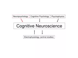
Cognitive Neuroscience is all about integrating information from diverse sources and levels……………..
Neuropsychology Neuroimaging Experimental cognitive psychology Theoretical models
2. Levels of Analysis in the understanding of semantic memory & semantic deficits? Gene Neurotransmitters(chemical)- psychopharmacology Neuron- micro (individual neuron), macro (networks) Functional brain areas – neuropsychology, neuroscience Computational modelling - cognitive science,psycholinguistics Behaviour – Neuropsychology & Normals, cognitive psychology, cognitive neuroscience. Social Interaction – Social Psychology: social cognition, Social Identity, Cultural – anthropology, socio-linguistics, cross-cultural psychology, social neuroscience.
Levels of Analysis? AD OL A N Hippocampal cell damage alcoholism
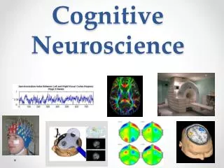
Areas activated during mental rotation exercise Brain areas associated with recognition of previously spoken words. Areas in yellow are associated with accurate recognition Areas in red are negatively correlated with accurate recognition
The left image is an averaged image of 10 nondrinker/social drinker young women, and the right is an averaged image of the 10 alcohol-dependent young women. Red, orange, and yellow show where the brain was active during spatial working memory, with yellow indicating the highest level of activity. Notice that there is less yellow in the back (bottom of the picture) right of the alcohol-dependent women's fMRI. Any Comment?
Brain-imaging level Control Mild Cognitive impairment Mild Alzheimer's Disease Any comment? [note prior to brain imaging autopsy only way to diagnose AD
3. Cautionary tales • fMRI • Alzheimer plaques & behaviour • Brain Lesions • Priming as tapping semantic memory
(i) Functional Neuroimaging • Permit the visualisation of memory processes in the healthy brain • Studies allow experimental designs to target specific memory processes
Functional Neuroimaging: Warning 1 • Signals are derived from local changes in blood flow, not neural activity. • Measures of neural activity are therefore indirect . • This means there is a limit to the temporal & spatial fidelity of activation. • In PET scans the local vascular changes affect the distribution of the injected radionuclide [O15 ] • In fMRI magnetic properties are blood-oxygen dependent
Functional Neuroimaging: Warning 2 • In the specification of mental processes signified by the activation there has to be a great deal of psychologicalinterpretation • This is usually done by activations arising from thedifferencebetween 2 tasks (subtraction method) • Differences are open to different interpretations • Differences are confounded by factors such as task difficulty or trial duration
Functional Neuroimaging: Warning 3 • Neuroimaging constraints: influence task design – need to block stimuli into homogeneous conditions – so that between conditioncomparisons can be made. • In fMRI each condition lasts for 30 sec • In PET each condition lasts for 2 min Rapid development of techniques at the present time attempting to combine the temporal resolution of ERP with the spatial resolution of fMRI
Functional Neuroimaging: Warning 4 • brain imaging techniques cannot compete with the psychological analysis of behaviour. • The thoughtfulness and accuracy of the psychological analysis of behaviour has now become even more important – as it feeds into neuroimaging studies. [e. g. ‘face recognition’ centre may turn out to be just ‘picture recognition’ centre: if use same photograph of the face!]
Physical ‘marks’ of Alzheimer's Disease Psychopharmacology Alzheimer's Disease Acetylcholine is required for the normal functioning of brain cells. As levels of the chemical decrease, plaques form, creating bundles of fibres (neurofibrillary tangles). Increase ventricle size
Psychopharmacology Alzheimer's Disease Rugg’s answer to the claim that those with high cognitive functioning have a particularly rapid AD decline. Incorrect - it is the opposite: the physical damage to the brain isn’t manifest in overt behaviour until the final stage in these patients – high cognitive functioning allows plasticity & flexibility to ‘cope’ better with AD. [New Scientist, 2005]
Physical expression is therefore not always apparent in behavioural expression
(iii) Brain Lesions • Provide the foundations of our knowledge about the biological organisation of human memory • Produce dramatic and unexpected deficits that provide clues about which brain regions are necessary for which brain processes.
Brain Lesions: warning 1 Behaviour of memory-impaired patients with brain lesions • does not delineate what process is subserved by the injured tissue • does reflect what uninjured brain regions can accomplish after the lesion.
Brain Lesions: warning 2 • Naturally occurring brain lesions often impair multiple brain systems by • direct insult to tissue • disconnection of interactive brain regions.
Brain Lesions: the problem • Therefore it is difficult to determine which deficit is a consequence of which part of a lesion
(iv) Behavioural studies • lexicon decision task Participants distinguish whether a word flashed up on the screen is a non-word (fuog) [no response] or a word (frog) [yes response]. The task is superficial and does not demand deeper semantic processing.
(iv) Behavioural studies • A prime is presented subliminally prior to the target word, reaction time to recognition of the target word is measured. • The prime word is either related (newt) or unrelated (book) to the target (frog). • Faster mean reaction time to targets preceded by related primes compared to unrelated primes are taken to show semantic priming.
As the task is nothing to do with word meaning semantic priming is considered to be tapping into the structure of semantic memory. BUT – warning can we assume this is automatic spreading activation within semantic memory?
Over a series of trials participants may become aware that there is some relationship between the prime and target and attempt to use an expectancy strategy • If this occurs then we are not measuring RTs that relate to automatic activation – a different memory process is involved in accomplishing the task – strategic retrieval. (more of this later)
Combination of methodologies • May overcome the limitations of each source of evidence • Provide powerful mutual constraints on ideas about memory systems • Lesion & Neuroimaging converge to identify neural networks & characterise processes
In normals activations for some memory tasks occur in brain regions that can be severely injured in neuropsychological cases and yet performance on a task may not be affected when the neuropsychological patient performs the memory task. • These activations may represent correlated memory processes that are not participating in the memory being measured in the specific neuroimaging study task. • Lesion evidence allows discrimination between activations that signify essential processes from non-essential processes for specific forms of memory being measured – impossible without lesion evidence.