Download
1 / 11
110 likes | 381 Views
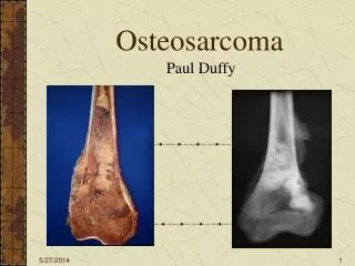
POST-OPERATIVE INFECTION AND INCREASED PATIENT SURVIVAL IN OSTEOSARCOMA : IS THERE A LINK? Lee Jeys, Rob Grimer, Simon Carter, Roger Tillman, Seggy Abudu, ROH, Birmingham, UK. Several known prognostic factors for osteosarcoma

E N D
POST-OPERATIVE INFECTION AND INCREASED PATIENT SURVIVAL IN OSTEOSARCOMA : IS THERE A LINK?Lee Jeys, Rob Grimer, Simon Carter, Roger Tillman, Seggy Abudu, ROH, Birmingham, UK • Several known prognostic factors for osteosarcoma • Lascelles showed increased survival in dogs with post-operative infection • Between 1966 & 2001 a consecutive series of 1264 patients underwent reconstruction for bone tumours • Reconstructions for metastases and benign excluded • Landmark analysis • Infections after 12 months excluded • Patients dying with 12 months excluded • Left 868 patients in study group
89/868 patients (10.3%) endured a deep infection within 12 months of EPR implantation • For osteosarcoma 10 year survival with no infection = 53.6% but with infection = 73.5% (p = 0.017). • Only significant with the diagnosis of osteosarcoma • Independent prognostic factor on cox regression analysis with other known factors with a HR=0.25 • No difference in percentage necrosis between groups, therefore, not due to more effective chemotherapy • Coley’s toxins (Streptococcus & Klebsiella) used historically • Supporting evidence for other tumours
89/868 patients (10.3%) endured a deep infection within 12 months of EPR implantation • For osteosarcoma 10 year survival with no infection = 53.6% but with infection = 73.5% (p = 0.017). • Only significant with the diagnosis of osteosarcoma • Independent prognostic factor on cox regression analysis with other known factors with a HR=0.25 • No difference in percentage necrosis between groups, therefore, not due to more effective chemotherapy • Coley’s toxins (Streptococcus & Klebsiella) used historically • Supporting evidence for other tumours
Aim Determine factors affecting local recurrence at proximal humerus Preservation of deltoid muscle Background Routine resection of deltoid muscle has been advocated by some authors Loss of deltoid could result in significant functional deficit, especially with newer forms of shoulder reconstruction Reverse shoulder prosthesis Allograft-prosthetic composite Methods 26 patients stage IIA or IIB OSA 12 females, 14 males Mean 19 yrs (range 7-47) Treatment Pre-op: DOX + IA-CDDP x 4 Limb-sparing surgery Post-op chemotherapy based upon tumor necrosis Determinants of Local Recurrence in Patients Undergoing Limb Sparing Surgery for Osteosarcoma of the Proximal HumerusPatrick Lin, Giri Gupta, Valerae Lewis, Christopher Cannon, Alan Yasko, UofTexas MDACC
Local Recurrence in Osteosarcoma of the Proximal Humerus • Results • 3/26 patients (11.5%) developed LR • 2 in sub-deltoid area • Tumor necrosis 50% & 70% • Both patients developed Mets & LR • Both patients died • 1 adjacent to chest wall (not sub-deltoid) • Pathologic fracture • 92% tumor necrosis • Alive 8 years after excision of LR • Conclusions • Local recurrence may depend on several factors, including margins, response to chemotherapy, and pathologic fracture • Routine resection of the deltoid muscle does not appear to be warranted
Radiation Induced Pathologic Fractures After Surgery for Soft Tissue Sarcomas • Aim of Study: • Determine healing rates of radiation induced fractures • Determine results of surgical management • Fracture Fixation vs Endoprosthetic Replacement • Background: • Previous studies established risk factors for fracture • Helmstedter (CORR 2001) - prophylactic IM nail with periosteal stripping • Lin (CORR 1998) - Consider primary arthroplasty in proximal & distal femur • Methods: • Retrospective review 1986 to present • 32 patients with 34 fractures (2 acetabular fractures - 1o THA) Kevan Saidi, Anthony Griffin, Peter Ferguson, Robert Bell, Jay Wunder, Mount Sinai Hospital, Toronto
Radiation Induced Pathologic Fractures After Surgery for Soft Tissue Sarcomas Results: 11 of 34 Fractures Healed (32%) • Femur: 25% (3/12) proximal 12% (1/8) diaphysis 50% (1/2) distal healed • Tibia: 100% (2/2) proximal 33% (1/3) diaphysis • Others: 60% (3/5) patella, metatarsals • 5/16 (31%) healed after periosteal stripping • 4/9 (44%) Men & 7/23 (30%) Women Healed • 2/8 (25%) healed after 50 Gy • 9/24 (38%) healed after 66 Gy • Risk Factors For Fracture (Holt, JBJS 2005): • Females > 50, High Dose Radiation (60 or 66 Gy), Proximal 2/3 Femur Conclusion: • Fractures of the proximal 2/3 of the femur and the tibia diaphysis are at high risk of non-union • Primary endoprosthetic replacement should be considered when treating pathologic radiation induced fractures of the proximal femur OR x 2 4.5 yrs 5 yrs
EXTRACORPOREALLY IRRADIATED AUTOGRAFTS IN LIMB SALVAGE SURGERYHow it can solve difficult reconstruction problems Hazem Wafa, MD; Robert Grimer, FRCS; Simon Carter, FRCS; Roger Tillman, FRCS; Adesgun Abudu; FRCS (orth) The Royal Orthopaedic Hospital, Birmingham, UK
Aim of the study: To evaluate the oncological and functional outcome of extracorporeally irradiated autografts as a reconstructive technique in limb salvage surgery. Material: • 28 patients (17 females, 11 males). • Median age 16 years (range 7-66). • 11 Ewing’s sarcoma, 8 osteogenic sarcoma, 4 parosteal osteosarcoma, 3 chondrosarcoma, 1 angiosarcoma, and 1 adamantinoma. • 14 pelvis, 6 tibia, 3 humerus, 2 scapula, 1 femur, 1 ulna, 1 metacarpal Methods: • Wide en-bloc resection of the lesion. • Removal of soft tissues and tumor off the excised bone segment. • Wrap in two layers with Vancomycin and pack in a sealed box. • Extracorporeal irradiation with 90 Gy (35 minutes). • Reimplantation of the irradiated autograft. • Primary hip replacement was used in 7 cases, pedicled vascularized fibular graft in 4, PMMA in 2, and non-vascularized fibula in 1.
Results: • The average follow-up 33.9 months. • 19 patients were alive (67.9%), 17 NED and 2 AWD. • Local recurrence in 3 cases (10.7%), all occurred in the soft tissues. • The average time to union was 7.7 months. • Deep infection developed in 2 cases (7.1%) necessitating graft removal. • Fracture of the graft occurred in 2 cases, graft resorption in 2, and non-union at 2 sites. • The mean MSTS functional score was 25.5 / 35 Conclusions: • Extracorporeal irradiation and re-implantation of bone is an excellent technique which can solve difficult reconstruction problems in appropriately selected patients. • This technique does not carry an increased risk of local recurrence and the overall results are comparable to other reconstructive techniques.