Download
1 / 64
670 likes | 696 Views
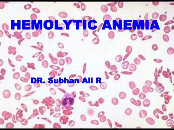
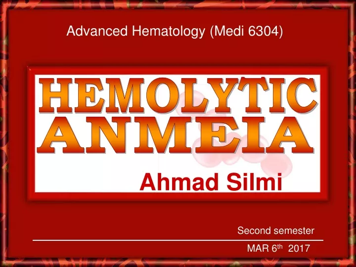
Advanced Hematology (Medi 6304). HEMOLYTIC. ANMEIA. Ahmad Silmi. Second semester MAR 6 th 2017. Hemolytic anemia: Definition

E N D
Advanced Hematology (Medi 6304) HEMOLYTIC ANMEIA Ahmad Silmi Second semester MAR 6th 2017
Hemolytic anemia: Definition • Anemia results from an excessive hemolysis of RBC’s • BM compensatory capacity: 6-8 folds of normal • Several folds destruction rate • Life span with erythrocytes less than 30 days • Anemia developed (Signs & symptoms) • Two type of hemolysis: • 1. Intra-vascular hemolysis • 2. Extra-vascular hemolysis
Hemolytic anemia: Hemolysis routes • Intravascular Hemolysis • Senescence: Reduced biological activities • Glycolytic pathway deterioration • - Loss of NADPH and NADH pool • - loss of ATP generation: • loss of Membrane permeability & deformability • Structural membrane abnormalities • Intracelluar Na+ () Intracelluar K+ () • Surface-volume ratio () Spheroidals h
Hemolytic anemia: Hemolysis routes • Intravascular Hemolysis (cont.) • Hemoglobin release into the circulation (Free) • Loaded on Haptoglobin (To BM or Liver) • Upon haptoglobin saturation • - Free Hemoglobin filtration (Kidney) & Reabsorption • - Excessive hemolysis • Reabsorption capacity exceeded • Hemoglobinuria and Hemoglobinemia
Hemolytic anemia: Hemolysis routes • Extravascular Hemolysis (cont.) • IgG accumulation on effete erythrocytes • RES Macrophages recognition & phagocytosis • Degradative enzymatic digestion by macrophages • Hemoglobin disassembling: • - Iron released Transferrin BM or Hepatocytes • - Globin components amino acids a.a. pool • - Protoporphyrin Bilirubin Conjugated (Liver) • Intestine (via Bile) • Stercobilinogen & Stercobilin (with feces) • Reabsorption to Kidney Urobilinogen (Urine
Hemolytic anemia: Clinical and diagnostic features • Associated with excessive destruction of erythrocytes • Liver capacity fail to conjugate • Bilirubin concentration elevated • Jaundice • Urobilinogen (in urine) elevated • Darkening of urine on standing • Stercobilinogen (in feces) elevated • Serum haptoglobin: absent
Hemolytic anemia: Complications Hyperbilirubinemia
Hemolytic anemia: Complications Hyperbilirubinemia
Hemolytic anemia: Complications Hyperbilirubinemia
Hemolytic anemia: Complications Hypercoagulability
Hemolytic anemia: Complications Hypercoagulability
Hemolytic anemia: Complications Hypercoagulability
Hemolytic anemia: Complications Hypercoagulability
Hemolytic anemia: Clinical and diagnostic features • CBC and Blood film examination
Hemolytic anemia: Diagnostic features
Hemolytic anemia: Clinical and diagnostic features • CBC and Blood film examination
Hemolytic anemia: Clinical and diagnostic features • CBC and Blood film examination
Hemolytic anemia: Clinical and diagnostic features • Associated with increased erythropoiesis rate: • Reticulocytosis and other evidences of BM hyperactivation
Hemolytic anemia: Clinical and diagnostic features • Associated with increased erythropoiesis rate: • Reticulocytosis and other evidences of BM hyperactivation
Hemolytic anemia: Clinical and diagnostic features • Associated with increased erythropoiesis rate: • Reticulocytosis and other evidences of BM hyperactivation
Hemolytic anemia: Clinical and diagnostic features • Associated with increased erythropoiesis rate: • BM index (Hyperplasia): Myeloid:Erthroid Hyperplasia (1:1)
Hemolytic anemia: Clinical and diagnostic features • Associated with morphological abnormalities: • Osmotic fragility and Autohemolysis • - Incubation of test RBC’s in hypotonic solution under sterile • conditions for 24 hours.
Hemolytic anemia: classification • Classification: Based on the etiology and pathophysiology • 1. Hereditary: 2. Acquired: • a. Membrane defects a. Immune-mediated: • (ex. Spherocytosis) Auto~ , Allo~ , Drug-induced ~ • b. Metabolic defects b. Microangipathic (MAHA) • (ex. G6PD deficiency) TTP, HUS, DIC • c. Haemoglobin Abnormalities c. Paroxysmal Noctunal Haemoglobinuria • (ex. Hb-S and Hb-C) d. Secondary: • Infections, Liver and renal disease
Hemolytic Anemia: Membrane defect > Vertical interactions stabilize the lipid bilayer. >> Deficiencies of spectrin, ankyrin, or band 3 protein causes decoupling of the lipid bilayer from the underlying skeletal lattice >>>subsequent membrane loss in the form of microvesicles. >>>>This leads to the formation of spherocytes (hereditary spherocytosis).
Hemolytic Anemia: Membrane defect > Horizontal interactions provide mechanical stability to the membrane. >> defects include abnormal spectrin heterodimer association to form tetramers and defective skeletal protein interactions of junctional complexes at the end of the spectrin tetramers (spectrin, actin, protein 4.1) >>> result in fragmentation of the red blood cell (schistocytosis).
Hereditary spherocytosis • HS Molecular Defects: Spectrin • - Spectrin deficiency is the most common protein alteration in HS. • - Not all spectrin defects lead to the formation of spherocytes. • - Mutations that affect the self-association of Spectrin heterodimers lead to hereditary elliptocytosis. • HS Molecular Defects: Ankyrin • - Patients with ankyrin defects have prominent spherocytosis without other morphologic defects. • - About 2/3 of patients have a combined defect of both ankyrin and spectrin.
Hereditary spherocytosis • HS Molecular Defects: Band 3 • - Generally milder. • - Many of the mutations affect the protein’s ability to be inserted into membranes • - All patients with band 3 deficiency have a proportional • decrease in protein 4.2 because band 3 is apparently needed for 4.2 stability. • HS Molecular Defects: Band 4.2 • - Protein 4.2 deficiency is associated with loss of band 3; and, in some cases, a loss of ankyrin. • - The morphology in these patients is variable and sometimes stomatocytes and ovalocytes are more common than spherocytes.
Hereditary spherocytosis: Diagnosis • Required criteria: • At least one sign of increased red cell destruction: • (ex. reticulocytosis, increased bilirubin, decreased haptoglobin, increased lactate dehydrogenase). • Increased spherocytes and anisocytosis on the blood film. • Increased red cell osmotic fragility. • Optional criteria • 1. Positive family history • 2. Splenomegaly • 3. Anemia (1/3 of HS patients are NOT anemic)
Hereditary ellipocytosis: - HE is inherited as an autosomal dominant gene. Heterozygous individuals are asymptomatic; homozygous individuals have mild to severe hemolysis. - Spectrin and band 4.1 mutations are invloved in the etiology of HE; abnormalities in protein 4.1 are much less common than spectrin mutations
Hereditary ellipocytosis: - The hallmark is the presence of large numbers of elliptocytes in the peripheral blood. - Red cell fragmentation may be seen in the more severe forms. - Osmotic fragility is generally normal. - If hemolysis is present, reticulocytosis, and other evidence of red cell destruction will be apparent as listed earlier for HS.
Hemolytic anemia: classification • Classification: Based on the etiology and pathophysiology • 1. Hereditary: 2. Acquired: • a. Membrane defects a. Immune-mediated: • (ex. Ellipocytosis, Spherocytosis) Auto~ , Allo~ , Drug-induced ~ • b. Metabolic defects b. Microangipathic (MAHA) • (ex. G6PD deficiency) TTP, HUS, DIC • c. Haemoglobin Abnormalities c. Paroxysmal Noctunal Haemoglobinuria • (ex. Hb-S and Hb-C) d. Secondary: • Infections, Liver and renal disease
Glucose 6-phosphate dehydrogenase (G6PD): G6PD Glucose G6P6-PG NADP NADPH Reductase Hexose Monophosphate Shunt GSH GSSH Peroxidase H202 H2O
G6PD deficiency: • G6PD gene is located on the X chromosome (q28). • Lyon’s hypothesis: • Heterozygous female: 2 cell populations • 50% of cell are G6PD normal & 50% are deficient • Clinically: features ranging from normal to severely deficient • G6PD variants: • - Point mutations (single amino acid substitution) • - >400 G6PD variants were identified. • - variants shows variable enzyme stability and/or kinetics
Chronic nonspherocytic hemolytic anemia (CNHA) • Evidenced by chronic hyperbilirubinemia, decreased serum haptoglobin level, and increased lactate dehydrogenase level. • The RBC morphology is unremarkable and is referred to as nonspherocytic. • The hemolysis is mostly extravascular (no hemoglobinurea). • Characterized by chronic anemia diagnosed at birth as having neonatal hyperbilirubinemia and the hemolysis continues into adulthood. • Usually with compensatory reticulocytosis but can be transfusion dependent.
G6PD deficiency: Clinical features • Normally, patients are asymptomatic till …. • Hemolytic crises: Intravascular hemolytic anemia following to the exposure of oxidizing agents • jaundice: 2-3 days after exposure • Anemia worsens till the 7th-8th day • Self limiting anemia • G6PD deficiency & Favism • G6PD deficiency and Resistance to P. falciparum
G6PD deficiency Pathogenesis
G6PD deficiency: Consequences Blister cells: red cells with a clearing at the periphery, like a blister Arises when bite cells undergo repair of the cell membrane, resulting in a clearing within the red cell where the Heinz body previous was. Bite cells: red cells with ‘bites’ taken out Bites’ are from splenic macrophages removing the part of the red blood cell with a Heinz body.
G6PD deficiency: Following to oxidative stress • Blood film morphology: • Normocytic, normochromic anemia • Red cells fragmentation ( Bite cells and Blister cells) • Occasional spherocytes • Dimorphisms (Females)
G6PD deficiency: Following to oxidative stress • Hemolytic features: • Increased reticulocytes after hemolytic crises • Hemoglobinemia & Hemoglobinuria