Download
1 / 64
690 likes | 1.16k Views
Rheumatoid Arthritis. DOM MR Week of 9/8/2008 Rozina Mithani. www.powerpointpresentationon.blogspot.com. Goals. General Approach to Arthritis Rheumatoid Arthritis Diagnostic Criteria Pathophysiology Therapeutic Approach Disease Severity and Course. Rheumatoid Arthritis: Definition.

E N D
Rheumatoid Arthritis DOM MR Week of 9/8/2008 Rozina Mithani www.powerpointpresentationon.blogspot.com
Goals • General Approach to Arthritis • Rheumatoid Arthritis • Diagnostic Criteria • Pathophysiology • Therapeutic Approach • Disease Severity and Course
Rheumatoid Arthritis: Definition • Progressive, systemic, inflammatory disorder • Unknown etiology • Characterized by • Symmetric synovitis • Joint erosions • Multisystem extra-articular manifestations
Epidemiology of Rheumatoid Arthritis • Approximately 1% of the total adult population is affected by RA • 40% to 60% with advanced (functional class IV) RA will: • Survive 5 years or less following diagnosis • Die 10-15 years earlier than expected
Joint Pain • most common symptom • Pain (arthralgia) vs. Inflammation (arthritis) • Inflammation: • heat, redness, pain, swelling, loss of function • inflammatory arthritis (RA, SLE) vs. pain syndrome (fibromyalgia)
Number of Joints Affected • Inflammatory vs. Non-Inflammatory
Monoarticular Crystal-induced Infection Reactive Arthritis Hemarthrosis OA: joint effusions Autoimmune disease Psoriasis, IBD, AS, Behçet's Oligo/Polyarticular Monoarticular causes RA SLE Viral infection B19 Acute Serum Sickness Untreated Crystal-induced Vasculidities Number of Joints Affected
Inflammatory: i.e. RA Generalized AM stiffness > 30 min Resolves with movement Classic signs of inflammation Non-Inflammatory: i.e. Osteoarthritis Localized AM stiffness < 30 min Inflammatory vs. Non-Inflammatory
Arthrocentesis • Confirm diagnoses • Differentiate between inflammatory & noninflammatory • Therapeutic/Adjunct to Antibiotics • Labs: • cell count w/diff • crystal analysis • Gram stain & Culture • WBC >2000/µL indicates inflammatory arthritis • Arthroscopy • Evaluate ligamentous & cartilaginous integrity • Biopsy • Infectioun: aspirate thick or loculated fluid
RA • Systemic inflammatory autoimmune disorder • ~1% of population • Onset: 52 years • 40-70 years of age • <60 - 3-5:1 female predominance
Genetics • Increased incidence among Pima & Chippewa Native American tribes (5%) • Genetic & Environmental • HLA-DRB1*0401 & HLA-DRB1*0404 • Increased risk • Increased joint damage • Increased joint surgery
Macrophages: Produce cytokines Cytokines (TNF-α) cause systemic features Release chemokines recruit PMNs into synovial fluid/membrane TNF-α & IL-1: Proliferation of T cells Activation of B cells Initiates proinflammatory/joint-damaging processes TH-1 cells: Mediate disease processes Activate B cells B cells: Release cytokines Plasma cells that produce Ab Osteoclasts: Bone erosion Juxta-articular & Systemic osteoporosis Immunology
Pathophysiology • Swelling of Synovial lining • Angiogenesis • Rapid division/growth of cells = Pannus • Synovial thickening/hyperplasia • Inflammatory vascularized tissue • Generation of Metalloproteinases • Cytokine release • Infiltration of leukocytes • Change in cell-surface adhesion molecules & cytokines • Destruction of bone & cartilage
Bottom Line • Proliferation • Destruction of joints • Disability
Disease Trigger • Subclinical vs. Viral trigger • Lab manifestations up to 10 yrs before clinical • RF & anti-CCP (anti–cyclic citrullinated peptide) Ab • Increased CRP subclinical inflammatory disease • ADLs: • > 50% of pts stop working w/i 5-10 years of disease onset • ~ 80% disabled to some degree > 20 years • Life expectancy: decreased by 3-18 years
Clinical Presentation • Gradual onset • Stiffness & Swelling • Intermittent or Migratory involvement • Extraarticular manifestations • Myalgia, fatigue, low-grade fever, wt loss, depression
Stiffness & Swelling • Pain with pressure to joint • Pain with movement of joint • Swelling due to hypertrophy • Effusion • Heat • Redness
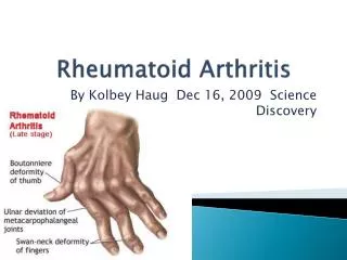
Physical Exam • Decreased grip strength • Boxing glove edema • Carpal tunnel • Ulnar deviation • Boutonniere/Swan neck deformities • Extensor tendon rupture
Anemia Rheumatoid nodules Pleuropericarditis Neuropathy Episcleritis, Scleritis Splenomegaly Sjogren’s Vasculitis Extraarticular Involvement
Seronegative polyarthritis Psoriatic arthritis Crystal-induced Tophaceous gout Pseudogout Erosive inflammatory OA Reiter’s Enteropathic arthritis SLE Paraneoplastic syndrome Differential
Diagnostic Criteria • Symmetric peripheral polyarthritis • AM Stiffness >1 hour • Rheumatoid nodules • Laboratory features • Radiographic bone erosions
Symmetric Peripheral Polyarthritis • 3 or more Joints for >6 weeks • Small Joints • Hands & Feet • Peripheral to Proximal • MCP and PIP Joints • SPARES DIP • MTP & Plantar subluxation • Leads to Deformity & Destruction of Joints • Erosion of cartilage and bone
Stiffness • AM or after Prolonged Inactivity • Bilateral • In/Around Joints • > 1 hours • Reflects severe joint inflammation • Better with movement • Present >6 weeks
Rheumatoid Nodules • Extensor surfaces • elbows • Very Specific • Only occur in ~30% • Late in Disease
Laboratory Features • RF • 70-80% of pts • Overlap with HCV/Cryoglobulinemia • Anti-Cyclic Citrulline Peptide (anti-CCP) • Rare overlap with HCV • Acute Phase reactants • ESR, CRP monitoring disease activity
Rheumatoid Factor • IgM against IgG • IgM+ pts: more severe disease & poorer outcome • Non-specific • SLE, Sjögren's, Sarcoidosis, Chronic infections
Anti-CCP • IgG against synovial membrane peptides damaged via inflammation • Value in IgM-RF negative • Sensitivity (65%) & Specificity (95%) • Predictive of Erosive Disease • Disease severity • Radiologic progression • Poor functional outcomes
Other Lab Abnormalities • AOCD • Thrombocytosis • Leukocytosis • ANA • 30-40% • Inflammatory synovial fluid • Hypoalbuminemia
Radiology • Evaluate disease activity & joint damage • Bony decalcification • Baseline AP views • Initiation of DMARDs
Radiological Studies • Plain Films • Bilateral hands & feet • Only 25% of lesions • Less expensive • Through bone cortex around joint margins • Color Doppler U/S & MRI • Early signs of damage i.e. Erosions • Bone Edema - even with normal findings on radiography
Mild Disease • Arthralgias • >3 inflamed joints • Mild functional limitation • Minimally elevated ESR & CRP • No erosions/cartilage loss • No extraarticular disease i.e. anemia
Moderate Disease • 6-20 Inflamed joints • Moderate functional limitation • Elevated ESR/CRP • Radiographic evidence of inflammation • No extraarticular disease
Severe Disease • >20 persistently inflamed joints • Rapid decline in functional capacity • Radiographic evidence of rapid progession of bony erosions & loss of cartilage • Extraarticular disease: • AOCD, Hypoalbuminemia
Prognostic Features • RF & Anti-CCP antibodies • Early development of multiple inflamed joints and joint erosions • Severe functional limitation • Female • HLA epitope presence • Lower socioeconomic status & Less education • Persistent joint inflammation for >12 weeks
CV Disease • Leading cause of death ~50% • 2x more likely to develop MI • chronic, inflammatory vascular burden premature atherosclerosis • MTX: elevated homocysteine levels • Control inflammatory process = Decreased atherosclerosis/morbidity • Lipid screening & treatment • Control of obesity, Hyperhomocystinemia, DM, HTN • ASA
Other diseases • 70% more likely to have a stroke • 70% higher risk for developing infection • Likely 2/2 treatment • 44x more likely to develop NHL
Staging • Early • <3 months • Established/Persistent • 6-12 months • End-stage • Significant joint destruction • Functional disability
Management • Early and aggressive disease control • Rheumatologist Referral • Early/Undiagnosed: NSAIDs, short course Corticosteroids • Late/Uncontrolled: DMARD therapy • depends on the presence or absence of joint damage, functional limitation, presence of predictive factors for poorer prognosis • Goals • achieve NED & inflammation • no treatment to resolve erosions once they occur
Non-Pharmacologic: Referral to PT/OT Evaluate ADLs Assistive devices/splints Weight loss Smoking cessation Pharmacologic: Anti-inflammatory Interrupt progression Development of erosions Joint space narrowing Therapy
Pharmacologic Therapy • Analgesics • NSAIDs • Glucocorticoids • SAARD/DMARD • Anticytokine therapy