Download
1 / 27
280 likes | 547 Views
Naming Skeletal Muscles. Direction of the muscle fibers Named in reference to an imaginary line Rectus – straight (parallel to line) Oblique – at a slant to the line Relative Size of the muscle Maximus – largest Minimus – smallest Longus - longest. Naming Skeletal Muscles.

E N D
Naming Skeletal Muscles • Direction of the muscle fibers • Named in reference to an imaginary line • Rectus – straight (parallel to line) • Oblique – at a slant to the line • Relative Size of the muscle • Maximus – largest • Minimus – smallest • Longus - longest
Naming Skeletal Muscles • Location of the muscle • Named for the bone with which they are associated • Number of origins • Bi – two origins • Tri – three • Quad - four
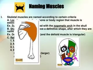
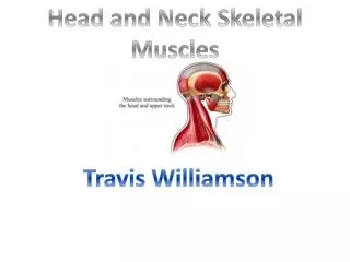
Naming Skeletal Muscles • Location of the muscle’s origin and insertion • Example is sternocleidomastoid = sternum, clavicle, and mastoid (process of the temporal bone) • Shape of the muscle • Deltoid means triangular • Action of the Muscle • Flexor, extensor, and adductor
Arrangement of Fascicles(Bundles of muscle fibers) Circular – concentric rings, used for external body organs that need to close by contracting. Also known as a sphincter Convergent – Converge to a single insertion tendon. Makes a fanlike or triangular muscle
Arrangement of Fascicles(Bundles of muscle fibers) • Parallel – length of fascicles run parallel to the long axis of the muscle • Fusiform – Type of parallel that results in a spindle-shaped muscle with an expanded belly • Pennate – feathered pattern. Short fascicles attach obliquely to a central tendon. • Unipennate attaches to one tendon • Bipennate attaches to two tendons • While multipennate attaches to manytendons
Facial Muscles Frontalis – Covers frontal bone. Allows you to raise your eyebrows and wrinkle your forehead Occipitalis – Covers occipital bone and pulls the scalp posteriorly Orbicularis Oculi – circular muscle around the eye which allows your eyes to close Orbicularis Oris – Circular muscle of the lips, also known as the kissing muscle Zygomaticus – Extends from the corner of the mouth to the cheek bone. Also known as the smiling muscle
Chewing Muscles Buccinator – Runs horizontally across the cheek and inserts into the orbicularis oris. It is also classified as a facial muscle. Responsible for flattening cheeks Masseter – covers the angle of the jaw bone. Responsible for closing the jaw by elevating the mandible Temporalis – fanshaped muscle that covers the temporal bone. It is the synergist to the masseter
Neck Muscles Platysma – single sheetlike muscle that covers the anterolateral neck. Produces a downward sag of the mouth Sternocleidomastoid – a pair of two headed muscles found on each side of the neck. If both contract, they flex the neck and lower your head. If one contracts, it rotates your head (Cranialaponeurosis - Sheetlike tendon on the top of the skull)
Trunk Muscles - Anterior PectoralisMajor – large fan-shaped muscle covering the upper part of the chest. It is used to adduct and flex the arm IntercostalMuscles – Deep muscles found between the ribs. They help with breathing by raising the ribcage.
Trunk Muscles – Abdominal Girdle Rectusabdominis – most superficial abdominal muscle. They flex the vertebral column, and compress the abdominal contents during defecation and childbirth Externaloblique – Make up lateral walls. They also flex the vertebral column, rotate the trunk, and bend it laterally Internaloblique – Deep to external, run at a right angle to them, and perform the same functions Transversusabdominis – Deepest abdominal muscle and it compress the abdominal contents
Trunk Muscles - Posterior Trapezuis – most superficial of these muscles and resembles a kite. They extend the head as well as elevate, depress, adduct, and stabilize the scapula LatissimusDorsi – Covers the lower back and is responsible for extending and adducting the humerus ErectorSpinae – It is composed of the longissimus, iliocostalis, and spinalis. These are the main extensors of the back as well as help control the action of bending over at the waist Deltoid – triangular shaped muscles that form the shape of the shoulder and are the prime movers of arm abduction
Muscles of the Upper Limb Bicepsbrachii – “The forearm muscle” that is the prime mover for flexion and is used to supinate the forearm Brachialis – muscle deep to the bicep that is used for elbow flexion Brachioradialis – a weak arm muscle Tricepsbrachii – the most powerful prime mover of elbow extension and is the antagonist of the bicep
Use the descriptions and your knowledge of the body’s muscles to label the figures on the back of this paper
Muscles of the Lower LimbMovement at the Hip GluteusMaximus – “Butt muscle” It is a powerful hip extensor, especially when power is needed (jumping and stair climbing) GluteusMedius – Deep to the maximus muscle and is a hip abductor and stabilizes the hip during walking Iliopsoas – a fused muscle (itiacus and psoasmajor) that is the prime mover of hip flexion and stabilizes upper body from falling backward while standing Adductormuscles – a group of muscles that adduct the hips together
Muscles of the Lower LimbMovement at the Knee Hamstring Group – Muscle mass of the posterior thigh that consists of the bicepsfemoris, semimembranosus, and the semitendinosus Sartorius – a weak thigh flexor which helps with crossing your legs Quadriceps Group – The muscles that powerfully extend the knee consists of the rectusfemoris and three vastus muscles
Muscles of the Lower LimbMovement at the Ankle and Foot TibialisAnterior – This dorsiflex and invert the foot ExtensorDigitorumLongus – it is the prime mover of toe extension and is also a dorsiflexor of the foot Fibularis Muscles – composed of the longus, brevis, and tertius. The group plantarflexes and everts the foot Gastrocnemius – “Calf muscle.” It is the prime mover for plantarflexion of the foot Soleus – also a strong plantar flexor
Use the descriptions and your knowledge of the body’s muscles to label the figures on the back of this paper Take 2
Demonstrate Each group will be assigned some muscles. You will have a few minutes to come up with some a “minute to win it” activity to help other feel the discussed muscles. Try not to use ones that waste a lot of materials (example: the tissue box with one hand)
The Ultimate “Test” I am going to show a video. As the video plays, I want your group to stand when your muscle group is being worked and sit when your muscles aren’t.