Download
1 / 23
320 likes | 1.04k Views
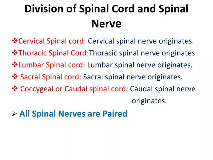
Division of Spinal Cord and Spinal Ner ve. Cervical Spinal cord: Cervical spinal nerve originates. Thoracic Spinal Cord: Thoracic spinal nerve originates Lumbar Spinal cord: Lumbar spinal nerve originates. Sacral Spinal cord: Sacral spinal nerve originates.

E N D
Division of Spinal Cord and Spinal Nerve Cervical Spinal cord:Cervical spinal nerve originates. Thoracic Spinal Cord:Thoracic spinal nerve originates Lumbar Spinal cord: Lumbar spinal nerve originates. Sacral Spinal cord: Sacral spinal nerve originates. Coccygeal or Caudal spinal cord: Caudal spinal nerve originates. All Spinal Nerves are Paired
Number of Spinal Nerves The spinal nerves are arranged in pairs. In goat e.g., there are usually thirty six (36) pairs of spinal nerves. 1. Cervical (C)nerves 8 pairs 2. Thoracic (T) nerves 13 pairs 3. Lumbar (L) nerves 6 pairs 4. Sacral nerves (S) 5 pairs 5. Coccygeal or Caudal nerves (Ca) : 4 pairs
Comparative Study in the Number of Spinal Nerves in Different animals and Birds Dog: 35-36 pairs Cat: 31 pairs Horse: 44 pairs Cattle: 36 pairs Pig: 39 pairs Bird (chicken): 30-33 pairs
Formation of Spinal Nerve Dorsal Root of Spinal Nerve (Sensory) Dorsal Root Ganglion Spinal Nerve. Pass through Intervertebral Foramina of Vertebral Column Ventral Root of Spinal Nerve (Motor)
Branches of a Spinal Nerve Dorsal branch of a spinal nerve which supplies to the epaxial muscle (muscle around the vertebral column) and skin. Ventral branch of a spinal nerve which supplies to the hypoaxial muscle (muscle and skin ventral to the transeverse process of Vertebral column. It also supplies to the fore and hind limb by forming brachial and lumbosacral plexus.)
Supply of Cervical Nerve • First and second nerve supply to the external ear, masseter muscle, muscle of the neck and throat region. • Third and fourth supply to the neck muscles. • Fifth, sixth and seventh ventral cervical nerve supply to the neck and in addition Phrenic nerve forms from these three nerves and supply to the diaphragm (in cat 4th to 7th). • Ventral branches of 6th to 8thforms brachial plexus and supply the fore limb.
Supply of Thoracic Nerve • Ventral branches of 1st one (in goat/sheep) or 1st and 2nd (in cattle, horse, dog) in association with last three ventral cervical spinal nerves forms brachial plexus and supply all the structures of fore limb. • Remaining ventral thoracic nerve (intercostal nerve) supply muscles in between ribs and skin. • Last ventral thoracic nerve supply in association with first lumber to the flank region.
Brachial Plexus Brachial plexus is formed by the anastomoses of the ventral branches of last three cervical and first 1st one (in goat/sheep) or 1st and 2nd (in cattle, horse, dog) ventral branches of the thoracicspinal nerves. Brachial plexus forms to supply all the structures of fore limb, and lateral wall of thorax and abdomen of animals.
Branches of Brachial Plexus Suprascapular n. Subscapular n. Axillary n. Pectoral n. Long thoracic n. Musculo- cutaneous n. Thoracodorsal n. Lateral Thoracic n. Ulnar n. Radial n. Median n. There are 11 pairs of nerves of brachial plexus
Supply of the branches of Brachial Plexus • Suprascapular nerve: Supraspinatus and infraspinatus muscles. Paralysis of this nerve causes “Sweeny”in horses which is characterized by the atrophy of the supra- and infraspinatus muscles. • Subscapular nerve: Supply to the subscapularis muscles. • Musculocutaneous nerve: Supply to the coracobrachialis muscle and biceps brachiimuscles. • Pectoral nerve: Supply to the pectoralis muscles. • Axillary nerve: Supply to the teres major, teres minor and deltoideus muscles.
Supply of the branches of Brachial Plexus 6. Radial nerve: Supply to the extensor muscles of the forearm. 7. Median and Ulnar nerve: Both supply to the flexor muscle. 8. Thoracodorsal nerve: Supply to the latissimusdorsi muscles. 9. Long thoracic nerve: Supply to the SerratusVentralis muscles. 10. Lateral Thoracic: Lateral aspect of the thoracic and abdominal region including skin.
Lumbar Spinal Nerve and Plexuses Supply of Lumbar nerves Iliohypogastric n. Skin of the abdomen and inguinal mammary gland. 2. Ilioinguinal n. Skin and muscles of abdomen, and inguinal mammary gland. Iliohypo- Gastric (L1) Femoral (L6) Ilioinguinal (L2) LareralCutaneous Femoral (L3) Genitofemoral (L4+5)
Lumbar Spinal Nerve and Plexuses Supply of Lumbar nerves 3.Lateral Cutaneous Femoral Nerve: Skin over the distal thigh and stifle joint. 4+5. GenitofemoralNerve: Inguinal mammary gland, Skin surrounding the vulva of cat and dog. Prepuce and scrotum of male Iliohypo- Gastric (L1) Femoral (L6) Ilioinguinal (L2) LareralCutaneous Femoral (L3) Genitofemoral (L4+5)
Lumbar Spinal Nerve and Plexuses Supply of Lumbar nerves 6.Femoral Nerve: Quadriceps muscle. Clinical problem: Paralysis Of this nerve cause fixation (stiffness) of Stifle joint causing whole limb incapable of Supporting weight. Iliohypo- Gastric (L1) Femoral (L6) Ilioinguinal (L2) LareralCutaneous Femoral (L3) Genitofemoral (L4+5)
Paravertebral Lumbar Anesthesia Between the last thoracic and L1 and in between L1 –L2, and L2- L3 Indication: Laparotomy: (Opening of the abdomen through flank* region). Most of the operation of the abdominal and pelvic organs. Operation of the mammary gland in female and genitalia of the male. * Soft lateral abdominal wall consisting of abdominal muscles, fascia, and skin.
Lumbosacral Plexus Definition: It is the anastomoses of the Ventral branches of last three lumbar and First two sacral nerves.
Lumbosacral Plexus Cranial Gluteal n. Caudal Gluteal n. Cranial Gluteal Nerve: supply to the middle and deep gluteal muscle. 2. Caudal Gluteal Nerve: supply to the Superficial gluteal muscle.
Lumbosacral Plexus 3.Caudal Cutaneous Femoral n. : Skin of the thigh and knee joint 3 • 4.Pudendal n. • In the male: Penile muscles and glans penis. • In the female: Vulva. • Both in male and female: skin around the anus. 4
Lumbosacral Plexus 5. Caudal Rectal n. : Caudal rectal part, sphincter of anus, and skin around the anus. 6. Sciatic n.: Largest and thickest of the lumbosacral plexus. Supply to the deep gluteal, obturator, Quadriceps, and gemelli muscle. Also to the capsule of the hip Joint.
Lumbosacral Plexus 7. Tibial nerve: Most of the flexor muscles of the leg. 8. Fibular nerve: Most of the extensor muscles of the leg. Sciatic Nerve Tibial Nerve Fibular Nerve
Sacral Spinal Nerve • Usually 5 pairs. • The first to forms the lumbosacral plexus. • The last 3 pairs innervates the rectum, anus and perineum (around the anus).
Epidural Anesthesia • Desensitization of part of the lumbosacral plexus, sacral, and caudal nerves by placing the local anesthetics on the epidural space through intervertebral foramina. • Two types of epidural anesthesia: (1) High epidural anesthesia (2) Low epidural anesthesia.
Epidural Anesthesia High epidural Low epidural Indication of epidural anesthesia: High epidural: Caesarian (Spell) section. Low epidural anesthesia: Docking and any operation in the perineum.