Download

1 / 19
210 likes | 524 Views
Single Umbilical Artery. Resmy Palliyil Gopi. Umbilical cord. 2 umbilical arteries 1 umbilical vein Rudimentary allantois Remnant of omphalomesenteric duct Whartons jelly. Incidence. Most common congenital malformation of the umbilical cord 5-10 in 1000 births (35-70 in 1000 twin births)

E N D
Single Umbilical Artery Resmy Palliyil Gopi
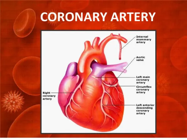
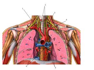
Umbilical cord • 2 umbilical arteries • 1 umbilical vein • Rudimentary allantois • Remnant of omphalomesenteric duct • Whartons jelly
Incidence • Most common congenital malformation of the umbilical cord • 5-10 in 1000 births (35-70 in 1000 twin births) • Left artery tends to be absent more often than the right • More common in Caucasian women
Conditions associated with single umbilical artery • Advanced maternal age • Multiparity • Maternal diabetes • Hypertension / toxemia • Ante partum hemorrhage • Poly/oligo hydramnios
Pathogenesis • Primary agenesis • Secondary atrophy or atresia of a previously normal umbilical artery • Persistence of the original allantoic artery of the body stalk
Associated prenatal complications • Isolated (1/2 to 2/3rd) • Associated congenital malformations (30%) • Fetal structural abnormalities • Chromosomal abnormalities (trisomy 18/13) • Genetic syndromes (VATER, sirenomelia sequence) • IUGR • Prematurity • Still birth and spontaneous abortions • Placental abnormalities
Diagnosis • prenatal • Ultrasound (only two vessels are seen and artery is abnormally large) • Color Doppler • postnatal • Examination of the cord
Management considerations • Detailed ultrasonographic examination • Fetal echocardiography • Fetal genetic testing • Enhanced fetal surveillance • Serial ultrasound to assess fetal growth • Biophysical profile, NST • Physical evaluation of the neonate • Neonatal renal ultrasonography
Prognosis - depends on the associated anomalies • Recurrence risk - Unknown
Abnormalities of the umbilical cord • Abnormalities of the cord length and diameter • Short cords and long cords
Abnormalities of the umbilical cord • Distortional abnormalities • loops, knots (true or false), torsions and twists True knot
Other abnormalities of the umbilical cord • Neoplasms of the umbilical cord • Thrombosis, hemangioma, hematoma, teratoma • edema