Download
1 / 36
500 likes | 2.01k Views
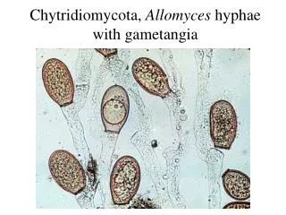
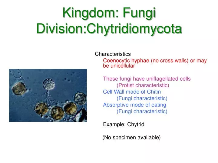
Kingdom: Fungi Division:Chytridiomycota. Characteristics Coenocytic hyphae (no cross walls) or may be unicellular These fungi have uniflagellated cells (Protist characteristic) Cell Wall made of Chitin (Fungi characteristic) Absorptive mode of eating (Fungi characteristic)

E N D
Kingdom: FungiDivision:Chytridiomycota Characteristics Coenocytic hyphae (no cross walls) or may be unicellular These fungi have uniflagellated cells (Protist characteristic) Cell Wall made of Chitin (Fungi characteristic) Absorptive mode of eating (Fungi characteristic) Example: Chytrid (No specimen available)
Kingdom: FungiDivision: Zygomycota Characteristics Coenocytic hyphae (no cell walls) Example: Rhizopus nigercans Observe the petri dish and slant of the living culture Rhizopus growing on agar. The white hairs are the haploid hyphae that make up the mycelium.
Kingdom: FungiDivision: Zygomycota Asexual Reproduction The long hairs under the scope are the hyphae that make up the mycelium. The mycelium can form sporangia, containing the asexually produced spores. The special hyphae bearing the sporangia are called sporangiophores.
Kingdom: FungiDivision: Zygomycota Sexual Reproduction Genetic recombination is by the process of conjugation which occurs when two strains grow close together. Each mycelium grows projections, called progametes. The cytoplasm of the two strains will fuse by a process called plasmogamy. At this point, the haploid nuclei pair off and the cell is said to be dikaryotic. The cell develops a rough, thick wall tha can protect the nucleus from harsh conditions. This structure is called a zygospore which than can go through karyogamy to form a diploid cell.
Kingdom: FungiDivision: Glomeromycetes Characteristics Coenocytic hyphae with mutualistic relationships with plant roots These fungi are called arbuscular mycorrhizae. The tips of the hyphae enter the plant roots and branch into tiny treelike structures called arbuscules. This division was formerly included in the zygomycetes but genetic evidence supports these should belong to a separate clade. Although there are only 160 species, they have a symbiotic association with 90% of all plant. (No specimen available)
Kingdom: FungiDivision: Ascomycota Characteristics Septate hyphae (cross walls) Asci Example: Peziza Observe the specimens of Peziza. The ascocarp is made of septate hyphae that is dikaryotic.
Kingdom: FungiDivision: Ascomycota Sexual Reproduction: The fruiting structure called an ascocarp is the result of sexual reproduction. The tips of the hyphae produce elongated sacs called asci. Within the asci, karyogamy occurs which produces a diploid nucleus. This nucleus divides by meiosis to create 4 haploid nuclei. The nuclei divide again by mitosis to form 8 haploid nuclei called ascospores. All the asci together are called the hymenial layer. Examine a prepared slide of Peziza, which shows a longitudinal cross section through the ascocarp.
Kingdom: FungiDivision: Basidiomycota Characteristics Septate hyphae (cross walls) Basidia Example: Coprinus, many others Observe the preserved specimens of club fungi. The examples are basidiocarps made up of dikaryotic hyphae. Examine living or preserved specimens of whole mushrooms.
Kingdom: FungiDivision: Basidiomycota Sexual Reproduction: The fruiting structure called a basidiocarpis the result of fusion of haploid hyphae. The fusion of haploid hyphae produce dikaryotic hyphae which make up the basidiocarp. The tips of the hyphae produce club shaped basidia. Within the basidia, karyogamy occurs which produces a diploid nucleus. This nucleus divides by meiosis to create 4 haploid nuclei. The 4 haploid nuclei move into appendages at the end of the hyphae called basidiospores.
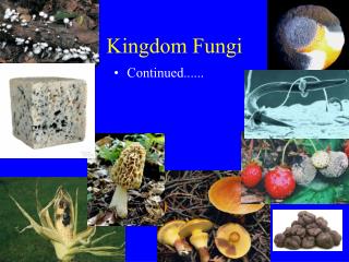
Kingdom: FungiDivision: Deuteromycota These species are called imperfect fungi because they don’t have (or we haven’t found) a sexual stage. All imperfect fungi reproduce asexually by means of conidia. Two examples of imperfect fungi are Penicillum notatum, which is used make the antibiotic penicillin, and Aspergillus niger, which is used to flavor foods. Examine living cultures of Penicillum notatum and Aspergillus niger. Note the coloring and texture of each culture.
Kingdom: FungiDivision: Deuteromycota Looking at prepared slides of the spore-bearing condidophores which house conidia. Spores of Penicillum appear blue-green and resemble a “kitchen fork”. Spores of Aspergillus appear black and resemble an “afro” hair style.
Lichens Lichens are actually a symbiotic relationship usually between a fungi and an algae. The fungal component is usually an ascomycota, but may be a basidiomycota. The fungus supplies moisture and shelter from high light intensity for the algae. The algae components are generally single-celled forms of green algae or cyanobacteria. The algae furnish food for the fungus. Lichens come in various colors and structures.
Kingdom: PlantaeDivision: Bryophyta Characteristics Nonvascular Seedless Example: Polytrichium Members of this division are small, gametophytes that usually grow upright. In moist places, they may form a large mat of vegetation. Individual plants consist of a stem-like stalk with attached leaf-like structures. Root-like rhizoids anchor the plant and absorb materials.
Protonema (Germinating Spores) The germinating structure is called a protonema. This structure is very similar to some filamentous green algae and is one piece of evidence that mosses might have evolved from some form of green algae. Be able to recognize this slide.
The Gametophyte Generation of a Moss The protonema have buds that develop into the “leafy” moss. Examine the moss plants provided for you. Male plants can be identified by the flower-like cluster of “leaves” at the tip of the gametophyte. In the female plants, the “leaves” closely surround the tip of the gametophyte.Be able to recognize the difference between male and female plants.
The Antheridium Examine the prepared slide of a longitudinal cross section of a male gametophyte tip. The antheridia contain sperm that are surrounded by sterile jacket cells. The antherdia are found in between paraphyses which are believed to protect the antheridia.
The Archegonium Examine the prepared slide of a longitudinal cross section of a female gametophyte tip. The archegonia has a swollen area called a venter which contains the egg. Above the venter is the neck of the archegonium. The archegonia are surrounded by paraphyses.
The Sporophyte Generation The sporophyte generation of a moss develops in the archegonium of a female gametophyte. A capsule develops on a long stalk called a seta. The capsue contains spores held inside by a hard covering called the operculum. A soft covering called a calyptra is part of the gametophyte generation and is created when the sporophyte grows out of the top of the female gametophyte.
The Sporophyte Capsule The sporophyte generation has photosynthetic tissue but is attached to the female gametophyte. It develops a capsule which is covered by a hard covering called an operculum. In the middle of the capsule is a structure called a columella that gives the capsule shape. Inside the capsule, spores are produced.
Kingdom: PlantaeDivision: Hepaticophyta Characteristics Nonvascular Seedless Example: Marchantia Members of this division are small, gametophytes that are usually found in two different types. 1. Thallose: flattened dorsoventrally 2. Leafy: resembles mosses Be able to recognize the examples seen at this station.
Liverwort Thallus The thallus is divided into an upper and lower secton. The upper level contains chlorophyll-bearing cells and is used for photosynthesis. Along the upper surface, there are pores that open up to air chambers that surround the chlorophyll-bearing cells used for gas exchange. The lower surface is divided into larger storage cells. Attached to the lower surface are rhizoids (single celled) and scales (multicellular) used for attachment and water absorption.
Antheridial and Archegonial Receptacles Liverworts produce gametangia on separate gametophytic plants. The male gametangia (the antheridia) resemble umbrellas and produce sperms. The female (the archegonia) resemble the spokes of a bicycle wheel and produce eggs.
Antheridial and Archegonial Receptacles The antheridium of a male liverwort contains sperm mother cells that produce sperm. The archegonium of a female liverwort contains a swollen area (venter) which holds the egg. The archegonium proceeds down into a neck with a canal that allows the sperm to get at the egg.
Liverwort Sporophyte The sporophyte generation (2n) is dependent on the gametophyte generation. It is attached to the female gametophyte by a foot and a small stalk called a seta. The capsule contains spores and elaters. The elaters are used for dispersal. They change with a chang in humidity and fling the spores away from the parent plant.
Asexual Reproduction Asexual reproduction occurs when the thallus produces gemmae cups that contain gemmae. The gemmae are dispersed when it rains and water splashes the gemmae out of the cup. This insures that the conditions are right for the gemmae to germinate and produce new plants.
Division Lycophyta Characteristics of a Club Moss These sporophytes have true stems, roots, and leaves. The stems are covered with small leaves called microphylls. They have modified leaves called sporophylls that bear sporangia. Be able to recognize the example Lycopodium in the jar.
Division Pterophyta(Psilophyta) Characteristics of a Whisk Fern The members of this group are unique among vascular plants in having sporophytes that lack true leaves or true roots. Be able to recognize the example Psilotum in the jar.
Division Pterophyta(Sphenophyta) Characteristics of a Horsetail These sporophytes have true stems, roots and leaves. These stems are covered with small leaves called microphylls that die at maturity. The stems are ribbed and contain silica in their inner cell walls. They have modified stems that produce small cones called strobili. Be able to recognize the example Equisetum.
Division Pterophyta Characteristics of a Fern The sporophytes has true stems, roots and leaves. The large leaves are called megaphylls or fronds. The fronds first appear tightly coiled and are called fiddleheads. This process is called circinate vernation. The specialized leaves with spores are called sporophylls. Be able to recognize the example Polypodium, Salvinia, etc.
Fern Frond Characteristics of a Fern Frond Ferns reproduce by producing spores. The fronds have small brown patches called sori. The sori are made up of clusters of sporangia which produce the spores. Many species have a protectiv covering called an indusium.
Fern Sporangia The sporangia have s conspicuous row of heavy-walled cells called an annulus. When the moisture in the cell changes, the annulus catapults spores out into the environment through the lip cells. The spores will develop into a prothallus.
Fern Prothallus The heart-shaped gametophyte generation of a fern is called a prothallus. Archegonia (which produce eggs) are usually found near the apical notch and antheridium (which produce sperm) are usually produced near the rhizoids (for absorption and anchorage). Eventually the sporophyte will grow out of the archegonia.
Fern Prothallus with a Sporophyte On the prothallus, only one zygote will develop into a sporophte. The simple structure consists of a small leaf, a root and a foot (the structure that attaches the sporophyte to the prothallus). The prothallus will eventually die off and the sporophyte generation will develop into a recognizable plant.