Download
1 / 37
400 likes | 599 Views
Neurotransmitter Types . Scanned from Squires et. al. Fundamental Neuroscience. Patton’s Requirements for Neurotransmitters.

E N D
Neurotransmitter Types Scanned from Squires et. al. Fundamental Neuroscience
Patton’s Requirements for Neurotransmitters • 1 Neurotransmitters must be synthesized in the presynaptic neuron: in the cell body and transmitted to the terminal end button or synthesized within the terminal end button.
2 The substance should be released from the nerve terminals in a chemically-pharmacologically identifiable form.
3 A neurotransmitter should reproduce at the postsynaptic cell the specific events (such as changes in membrane properties) that are seen after stimulation of the presynaptic neuron.
4 (A) The effects of a putative neurotransmitter should be blocked by competitive antagonists of the receptor for the transmitter in a dose dependent manner. • (B) Treatments that inhibit synthesis of the transmitter should block the effects of presynaptic stimulation
5. There should be active mechanisms to terminate the action of the putative neurotransmitter (uptake by the presynaptic neuron or glial cells through specific transport mechanisms, or enzymatic inactivation of the chemical messenger).
Two Large Categories of Neurotransmitters • Traditional • Nontraditional
Nontraditional synapses • Peptide synapses • Nitric oxide (the gas)
Ionotrophic synapses • nACh receptor is opened by ACH, such that all small ions (Na+, K+, Cl-) run down their diffusion gradiant.
Metatrophic • Neurotransmitter opens gates if coupled with G proteins.
Acetylcholine neuromuscular Junction • At the end of the 1930s and early 40s the general conclusion was reached that the first post synaptic event was a negative going depolarization of the muscle fiber. • Named the end-plate potential • Was restricted to the end-plate region. • Is sufficient in size to generate and action-potential which propagates the length of the fiber.
Acetylcholine • Dale investigated a substance obtained from ergot a fungal infection of cereal. • Slowed heart rate of the cat, and lowered blood pressure. • Hypothesized that acetylcholine occurred naturally in the body and hydrolized by an enzyme in the body accounting for the inpossabillity of it being isolate in from the body.
Lowe’s study on frog heart • Found that stimulation of the Vagus nerve caused heart1 to lengthen the beat to beat time frame slowing heart rate. • An aliquot of the perfusion fluid of heart1 when transfered to heart 2, with no stimulation of heart2’s Vagus nerve caused heart 2 lengthen its beat to beat time frame and slow heart 2 down.
Dale and Dudley’s study • Isolated acetylcholine from the spleen of cows and horses. • Demonstration that acetylcholine could be isolated from the ganglia of the sympathetic nervous system and at nerve terminals in the motor muscles of the sckelatal. system
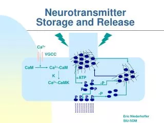
Construction-Destruction • Construction • Acetyl Co A + choline == Acetylcholine • Destruction • Acetylcholine + acetylcholinesterase == • choline + acetic acid
Fatt and Katz studyArrangement for Intracellular recording of post synaptic neuromuscular junction
Using the apparatus from the previous slide for Intracellular recording of post synaptic neuromuscular junction. The electrode is rode is progressively move away from the axon in mm steps
Electrical stimulation of the axon of the presynaptic membrane is different in from direct stimulation of the muscle fiber.
Iontophertic injections of Ach, developed by Nastuk (1953) further developed by Katz and del Catillo (1955)
Miniature end-plate potentials • In the resting membrane, small potentials of about 0.5mV can be recorded. • Could only be found at the end-plate in curarized preparations. • Could not be found in preparations where the muscle had been previously denervated two weeks previously.
Characteristics of the miniature end-plate potential • According to Katz, the frequency of the miniature end-plate potentials is controlled by the conditions of the presynaptic membrane, while the amplitude is controlled by the properties of the postsynaptic membrane
Quantal release of Acetylcholine • A combination of Mg and Ca can be used to block the release of ACH. • When this concentration if used appropriately, the size of the successive EPP will fluctuate in a step-wize manner (see curve below).