Download
1 / 1
10 likes | 218 Views
Phase unwrapping. Goal Accelerate the performance of the minimum L P Norm phase unwrapping algorithm using Field Programmable Gate Arrays (FPGAs). Figure 1: Wrapped Phase. Bio-Med. Enviro-Civil. S2. S3. S1. S4. S5. UNWRAP. L3. Validating TestBEDs. L2. R2. Fundamental Science. R1.

E N D
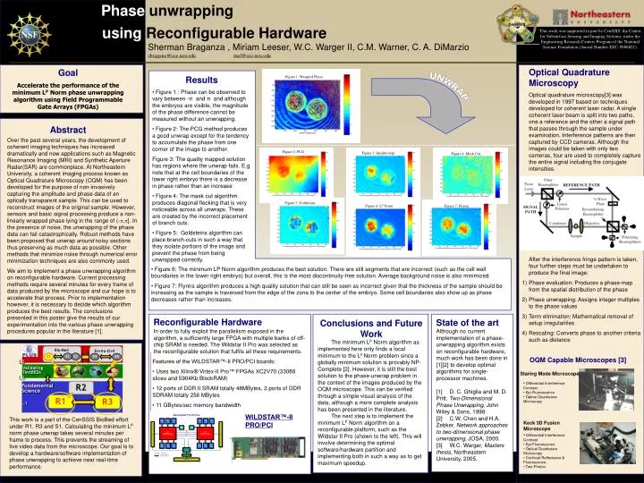
Phase unwrapping Goal Accelerate the performance of the minimum LP Norm phase unwrapping algorithm using Field Programmable Gate Arrays (FPGAs) Figure 1: Wrapped Phase Bio-Med Enviro-Civil S2 S3 S1 S4 S5 UNWRAP L3 ValidatingTestBEDs L2 R2 FundamentalScience R1 R3 L1 Abstract Over the past several years, the development of coherent imaging techniques has increased dramatically and now applications such as Magnetic Resonance Imaging (MRI) and Synthetic Aperture Radar(SAR) are commonplace. At Northeastern University, a coherent imaging process known as Optical Quadrature Microscopy (OQM) has been developed for the purpose of non-invasively capturing the amplitude and phase data of an optically transparent sample. This can be used to reconstruct images of the original sample. However, sensors and basic signal processing produce a non-linearly wrapped phase lying in the range of (-,]. In the presence of noise, the unwrapping of the phase data can fail catastrophically. Robust methods have been proposed that unwrap around noisy sections thus preserving as much data as possible. Other methods that minimize noise through numerical error minimization techniques are also commonly used. We aim to implement a phase unwrapping algorithm on reconfigurable hardware. Current processing methods require several minutes for every frame of data produced by the microscope and our hope is to accelerate that process. Prior to implementation however, it is necessary to decide which algorithm produces the best results. The conclusions presented in this poster give the results of our experimentation into the various phase unwrapping procedures popular in the literature [1]. Figure 2: PCG Figure 3: Quality map Figure 4: Mask Cut Figure 5: Goldsteins Figure 6: LP Norm Figure 7: Flynns using Reconfigurable Hardware This work was supported in part by CenSSIS, the Center for Subsurface Sensing and Imaging Systems, under the Engineering Research Centers Program of the National Science Foundation (Award Number EEC-9986821). Sherman Braganza , Miriam Leeser, W.C. Warger II, C.M. Warner, C. A. DiMarzio sbraganz@ece.neu.edumel@ece.neu.edu • Optical Quadrature Microscopy • Optical quadrature microscopy[3] was developed in 1997 based on techniques developed for coherent laser radar. A single coherent laser beam is split into two paths, one a reference and the other a signal path that passes through the sample under examination. Interference patterns are then captured by CCD cameras. Although the images could be taken with only two cameras, four are used to completely capture the entire signal including the conjugate intensities. • After the interference fringe pattern is taken, four further steps must be undertaken to produce the final image: • Phase evaluation: Produces a phase-map from the spatial distribution of the phase • Phase unwrapping: Assigns integer multiples to the phase values • Term elimination: Mathematical removal of setup irregularities • Rescaling: Converts phase to another criteria such as distance Results • Figure 1 : Phase can be observed to vary between -π and π and although the embryos are visible, the magnitude of the phase difference cannot be measured without an unwrapping. • Figure 2: The PCG method produces a good unwrap except for the tendency to accumulate the phase from one corner of the image to another. Figure 3: The quality mapped solution has regions where the unwrap fails. E,g note that at the cell boundaries of the lower right embryo there is a decrease in phase rather than an increase • Figure 4: The mask cut algorithm produces diagonal flecking that is very noticeable across all unwraps. These are created by the incorrect placement of branch cuts. • Figure 5: Goldsteins algorithm can place branch-cuts in such a way that they isolate portions of the image and prevent the phase from being unwrapped correctly. • Figure 6: The minimum LP Norm algorithm produces the best solution. There are still segments that are incorrect (such as the cell wall boundaries in the lower right embryo) but overall, this is the most discontinuity-free solution. Average background noise is also minimized. • Figure 7: Flynns algorithm produces a high quality solution that can still be seen as incorrect given that the thickness of the sample should be increasing as the sample is traversed from the edge of the zona to the center of the embryo. Some cell boundaries also show up as phase decreases rather than increases. Reconfigurable Hardware Conclusions and Future Work State of the art Although no current implementation of a phase-unwrapping algorithm exists on reconfigurable hardware, much work has been done in [1][2] to develop optimal algorithms for single-processor machines. [1] D. C. Ghiglia and M. D. Pritt, Two-Dimensional Phase Unwrapping, John Wiley & Sons, 1998. [2] C.W. Chen and H.A. Zebker, Network approaches to two-dimensional phase unwrapping, JOSA, 2000. [3] W.C. Warger, Masters thesis, Northeastern University, 2005. In order to fully exploit the parallelism exposed in the algorithm, a sufficiently large FPGA with multiple banks of off-chip SRAM is needed. The Wildstar II Pro was selected as the reconfigurable solution that fulfils all these requirements. The minimum LP Norm algorithm as implemented here only finds a local minimum to the L0 Norm problem since a globally minimum solution is provably NP-Complete [2]. However, it is still the best solution to the phase-unwrap problem in the context of the images produced by the OQM microscope. This can be verified through a simple visual analysis of the data, although a more complete analysis has been presented in the literature. The next step is to implement the minimum LP Norm algorithm on a reconfigurable platform, such as the Wildstar II Pro (shown to the left). This will involve determining the optimal software/hardware partition and implementing both in such a way as to get maximum speedup. OQM Capable Microscopes [3] • Features of the WILDSTAR™-II PRO/PCI boards: • Uses two Xilinx® Virtex-II Pro™ FPGAs XC2V70 (33088 slices and 5904Kb BlockRAM) • 12 ports of DDR II SRAM totally 48MBytes, 2 ports of DDR SDRAM totally 256 MBytes • 11 GBytes/sec memory bandwidth Staring Mode Microscope • Differential Interference Contrast • Epi-Fluorescence • Optical Quadrature Microscopy WILDSTAR™-II PRO/PCI This work is a part of the CenSSIS BioBed effort under R1, R3 and S1. Calculating the minimum LP norm phase unwrap takes several minutes per frame to process. This prevents the streaming of live video data from the microscope. Our goal is to develop a hardware/software implementation of phase unwrapping to achieve near real-time performance. Keck 3D Fusion Microscope • Differential Interference Contrast • Epi-Fluorescence • Optical Quadrature Microscopy • Confocal Reflectance & Fluorescence • Two-Photon


![[1]. D. O. Hogenboom, C. A. DiMarzio, T. J. Gaudette, A. J. Devaney, and S. C.](https://cdn2.slideserve.com/4449989/slide1-dt.jpg)