Download
1 / 1
10 likes | 83 Views
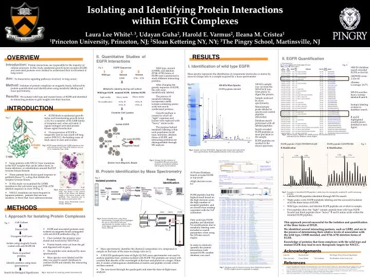
Isolating and Identifying Protein Interactions within EGFR Complexes Laura Lee White 1, 3 , Udayan Guha 2 , Harold E. Varmus 2 , Ileana M. Cristea 1 1 Princeton University, Princeton, NJ; 2 Sloan Kettering NY, NY; 3 The Pingry School, Martinsville, NJ. EGFR peptide GLWIPEGEK K Modification.

E N D
Isolating and Identifying Protein Interactions within EGFR Complexes Laura Lee White1, 3, Udayan Guha2, Harold E. Varmus2, Ileana M. Cristea1 1Princeton University, Princeton, NJ; 2Sloan Kettering NY, NY; 3The Pingry School, Martinsville, NJ EGFR peptide GLWIPEGEK K Modification EGFR peptide LTQLGTFEDHFLSLQR R Modification Fig. 9 EGFR 1 4 Da 4 Da 6 Da 4 Da Ab (anti-EGFR) EGFR 2 Magnetic bead EGFR 3 Cell Culture Freeze Cells Cryogenic Cell Lysis Isolate using magnetic beads coated with anti-EGFR Ab Elute co-purified proteins Identify proteins using mass spectrometry Search for Biological Significance Fig. 3 RESULTS OVERVIEW II. Quantitative Studies of EGFR Interactions II. EGFR Quantification Introduction: Protein interactions are responsible for the majority of cellular processes. In this study, epidermal growth factor receptor (EGFR) and associated proteins were isolated to understand their involvement in lung cancer. Aim: To characterize signaling pathways involved in lung cancer. Method:Isolation of protein complexes on magnetic beads, followed by protein quantification and identification using metabolic labeling and mass spectrometry. Results: We isolated wild type and mutant forms of EGFR and identified its interacting proteins to gain insights into their function. • MS-Fit database search revealed EGFR as first hit. • MOWSE score: 2.33e+10 • Protein Coverage: 24.7% MS-Fit searches heavy isotope labeled R and K. Isotopic labeling evident in database search. R and K highlighted modification’s detailed in next figure. I. Identification of wild type EGFR Fig. 8 EGFR Sequences Wild type Mutated Deletion L858R E746 – A750 Metabolic Labeling during cell culture Combine Cell Lysates Isolate EGFR Elution from Magnetic Beads Fig. 4 Wild type, mutant (L858R), and deletion (E746-A750) forms of EGFR were constructed to study different interacting proteins. After changing the genetic sequence of EGFR, the cells were metabolically labeled. The process of metabolic labeling incorporates stable isotope-containing amino acids into newly synthesized proteins. Growth medium is created in which all “light” arginines and lysines were replaced with “heavy” amino acids. The purpose behind metabolic labeling is that each population (wild type, mutation, deletion forms of EGFR, and interacting proteins) are distinguishable through MS analysis. 1 2 3 Mass spectra represents the distribution of components (molecules or atoms) by mass-to-charge ratio in a sample acquired by a mass spectrometer Fig. 6 100 kDa sample was cut from the SDS-PAGE into 1mm pieces to digest the protein. Sample analyzed by mass spectrometry. Monoisotopic peaks labeled in spectra and control peptides eliminated. Database search performed with all labeled peptides. Search revealed EGFR peptides as most prevalent in the sample. EGFR peptides are marked in the shown spectrum. 100 kDa Mass Spectra EGFR peptides labeled kDa Wild type EGFR mutated EGFR deletion EGFR 250 Heavy media Light media Heavy media 150 No modification +6 Da R +4 Da K +4 Da R +4 Da K 100 INTRODUCTION 75 • EGFR binds to epidermal growth factor and transforming growth factor α [1]. It is a member of the ErbB family of receptors and, when activated, it signals mitogen-activated protein kinase signal transduction. • Overexpression of EGFR is frequently seen in non-small cell lung cancer (NSCLC), the leading cause of fatal cancer in men and women. Fig. 1 50 37 Fig 8. MS-Fit results. Highest hit, EGFR protein. The modifications are indicated by the database search engine. 25 15 Fig 1. EGFR image labeled with L858R mutation in the activation loop and E746-A750 deletion in exon 19. (J. Guillermo Paez, 2004). Fig 6. Sample cut from SDS-PAGE, digested with trypsin and analyzed by mass spectrometry; EGFR peptides labeled on the mass spectrum. Fig. 2 Fig 4. Strategy for the isolation of mutated EGFR and associated proteins via metabolic labeling and magnetic bead isolation Fig. 7 • Some patients with NSCLC have mutations in the EGF receptor that can be either short, in-frame deletions, or substitutions around the EGF tyrosine kinase domain. • These patients have shown good response to gefitinib (Iressa™), a drug that inhibits the receptor tyrosine kinase. • In this study we examined the L858R mutation in the activation loop and E746-A750 deletion sequence in exon 19 (Fig. 1). • NSCLC mutations are most frequent in Japanese patients, patients that are non-smokers, or those that have adenocarcinoma. • X-Proteo Database Search revealed EGFR as top result • High decision score: 8.0 EGFR peptides had the highest result based on the high decision score, the high number of assigned peptides, and an overall error reading consistent with the ToF calibration. Only wild type EGFR peptides are identified. No modifications due to metabolic labeling were included as search parameters when using the XProteo search engine. In order to relatively quantify the protein interactions with mutant EGFR, another database was used. III. Protein Identification by Mass Spectrometry Fig. 5 Protein Identification Isolated proteins Fig 2. Images of adenocarcinoma of the lung. Top image is the control image of the tumor cell. Bottom image is the tumor treated with gefitinib. This treatment led to the death of most tumor cells (image from Weisenthal Cancer Group, 2006 [2]). Fig 9. Examples of identified EGFR peptides. Labels show the isotopically modified R- and K-containing peptides. • Labeled EGFR peptides identified through MS-Fit search. • Triple peaks verify EGFR metabolic labeling and the successful isolation of all the three forms of EGFR. • Wild type, mutation, and deletion EGFR peptides are evident in sample. • First peptides show the “light” isotopic peptide from wild type EGFR. Second and third peptides show “heavy” R and K amino acids within the mutated EGFR peptides. METHODS I. Approach for Isolating Protein Complexes Fig 5. Protein Identification using Mass Spectrometry. Example shown highlights the use of a MALDI Q-ToF mass spectrometer for protein analysis. Parts of the figure adapted from Steen and Mann, 2004 [4]. This approach proved successful for the isolation and quantification of the three forms of EFGR. We identified several interacting partners, such as GRB2, and are in the process of determining their relative levels of association with the wild type, L858R mutated, and E746-A750 deletion forms of EGFR. Knowledge of proteins that form complexes with the wild type and mutant EGFR may lead to new therapeutic targets for NSCLC. • EGFR and associated proteins were isolated on magnetic beads conjugated with anti-EGFR antibodies (Fig. 3). • Once isolated, the proteins were eluted and resolved by SDS-PAGE. • Protein bands were cut from the gel and digested with trypsin. • The peptides were analyzed by mass spectrometry. • Mass spectra were labeled and the data was used to search databases to confirm EGFR and identify the interacting partners. • Mass spectrometry identifies the chemical composition of a compound or sample on the basis of the mass-to-charge ratio (m/z). • A MALDI quadrupole time-of-flight (Q-ToF) mass spectrometer was used to analyze peptides from proteins isolated with EGFR. The peptides are mixed with a matrix that absorbs at the wavelength of the laser and placed on a target. When dry, it creates a heterogenous distribution of matrix and peptide crystals which are then ionized. • The ions travel through the quadrupole and enter the time-of-flight mass analyzer. Acknowledgements Luke De Bonnie Kaiser The Pingry School Science Department Anna Arnaudo Chase Palisch Val Carabetta The Members of the Cristea Lab References Udayan Guha, Raghothama Chaerkady, Arivusudar Marimuthu, A. Scott Patterson, Manoj K. Kashyap, H. C. Harsha, Mitsuo Sato, Joel S. Bader, Alex E. Lash, John D. Minna, Akhilesh Pandey. Comparisons of tyrosine phosphorylated proteins in cells expressing lung cancer-specific alleles of EGFR and KRAS. (2008)PMAS, in print. Larry Wiesenthal, M.D., P.h.D., Wiesenthal Cancer Group.(2006). Test identifies patients who benefit from targeted cancer drugs. Europeoan Hospital Journal. Ileana M. Cristea, John-William N. Carroll, Michael P. Rout, Charles M. Rice, Bran T. Chait, and Margaret ‘R. MacDonald., (2001). Tracking and Elucidating Alphavirus-Host Protein interactions. Joournal of Biological Chemistry. Volume 281, 40. 30269-30278 Hanno Steen, Matthias Mann.. The ABC’S (and XYZ’S) of Peptide Sequencing. (2004) Nature Reviews. September. Vol 5, 699-711 Fig 7. Database search of known masses from labeled mass spectra. EGFR is highest match of peptides in sample. Fig 3. Approach for studying protein interactions [3]