Download
1 / 38
420 likes | 576 Views
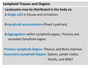
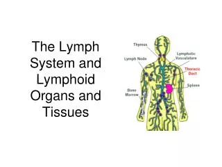
Immune Cells , Receptors, and Markers. Lymphoid Tissues and Organs. White blood cells or leukocytes serve as defenders against infection. They move around the body via the lymphatic and blood circulatory systems, and can leave and reenter the circulation. N.

E N D
Immune Cells , Receptors, and Markers.Lymphoid Tissues and Organs.
White blood cells or leukocytes serve as defenders against infection. • They move around the body via the lymphatic and blood circulatory systems, and can leave and reenter the circulation.
N • Leukocytes are classified according to their morphology, origin, and function into the following groups: • Cells of Myeloid lineage • Granular leukocytes. • Agranular leukocytes. • Cells of Lymphoid lineage • Granular leukocytes. • Agranularleukocytes.
Cells of Myeloid lineage: • - Granularleukocytes: Neutrophils, Eosinophils & Basophils. • - Agranular leukocytes: Monocytes , and macrophages. • Cells of Lymphoid lineage: • - Granular leukocytes: NK cell. • - Agranular leukocytes: T cell, B cell.
N • Cells of Myeloid lineage: • Granular leukocytes: • Neutrophils or polymorphonuclear cells: • ~60% of the peripheral blood leukocytes. • Very effective at killing bacteria. • Elevated number: mainly in acutebacterial infection.
Eosinophils: • Eosin-loving granules. • form 0-5% of WBCs. • Activated in parasitic helminthesinfectionand in allergic conditions.
N • Basophils: • Acidic cytoplasmic granules contain histamine. • Form 0-1% of WBCs. • Activated in allergicconditions.
N • Agranular leukocytes: • Monocytes , and macrophages: • -Monocytes in the circulation, and macrophage in the tissues. • 5-7% of peripheral blood leukocytes. • Scavenger cells for innate immunity. • Antigen presenting cells (APC).
Cells of Lymphoid Lineage: • Lymphocytes: • Account for ≈ 40% of WBCs. • According to type of receptor, and organs of differentiation (where they undergo basic training), Lymphocytes are classified into: • Thymus-derived cells (T Cells) • Bone marrow-derived cells (B Cells) • Natural killer cells (NK-Cells)
Thymus-derived cells: T lymphocytes: • Arise from Bone marrow • Enter the circulation from Thymus. • Identified by presence of CD3complex with T cell receptor (TCR).
Two subsets of T lymphocytes: • CD8 ⁺ T cells (cytotoxic): Restricted to the recognition of major histocompatibility complex (MHC) class I complexes • CD4⁺ T cells (helper): Restricted to the recognition of major histocompatibility complex (MHC) class II.
Major histocompatibility (MHC) molecules: • MHC class I molecule: found on all nucleated cells. Present antigens to CD8 T cells • MHC class II molecule: Present antigens to CD4 T cells • T cells which encounter antigen differentiate into effector T cells.
N • Bone Marrow derived cells: B cells: • develop within bone marrowand produce Igs. • IgM and IgD both are present on cells surface. • Isotype switching according to the type of infection. • Markers : CD19, and CD20.
N • Plasma cells: • - Terminally differentiated B cells. • - Immunoglobulin producing cells.
Natural killer cells (NK-Cells): • Forms 5-10% peripheral blood lymphocytes. • Lack both T and B cell receptors. • Very active in killing virallyinfected cells and tumor cells. • Have 2 receptors: • Killing activation receptor (KAR) • Killing inhibition receptor (KIR)
Normal cell Malignant or virally infected cell
Immune Cells Receptors: • Immune system depends upon receptors to distinguish own cells& organs (Self) from those of foreign origin (non self). • The immune cell receptors are classified into two types: • Preformed receptors. • Somatically generated receptors (epitope specific).
Preformed receptors. • Pattern recognition receptors (PRR). • Toll-like receptors. • The complement receptors. • The immunoglobulins Fc receptors. • The Natural Killer cell receptors.
Preformed Receptors • Pattern recognition receptors: recognize pathogen associated molecular patterns (PAMP). • Toll-like receptors: also activated by binding to a PAMP.
The Complement Receptors: • C3b (opsonin) fragments bind to its receptors on Phagocytes and B cells.
The immunoglobulins Fc receptors: -Antigen-antibody complex are recognized by Fc receptors on phagocytic cells. -The Fc receptor on mast cells that binds IgE is exceptional (bounded before interaction with the antigen).
N The Natural Killer Cell Receptors: Killer activation receptors (KARS): Recognize certain type of molecules expressed by unhealthy host cells. Killer inhibition receptors(KIRS): Recognize the presence of MHC class I , which is normally displayed on the host cell surfaces.
N • Somatically Generated Receptors: • B cell receptors (BCR): • Immunoglobulins (IgM &IgD) serve as B-cell receptors. • T cell receptors (TCR): • Cluster of differentiation 3 (CD3) and TCR bind to MHC. • CD4and CD8 are co-receptors.