Download
1 / 1
10 likes | 84 Views
Developed pipeline for longitudinal brain image analysis to study neurodegenerative diseases, improving accuracy & efficiency.

E N D
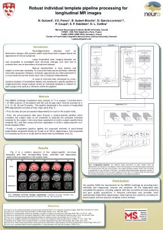
Robust individual template pipeline processing for longitudinal MR images N. Guizard1, V.S. Fonov1, B. Aubert-Broche1, D. García-Lorenzo1,2, P. Coupé3, S. F. Eskilden4, D. L. Collins1 1Montreal Neurological Institute, McGill University, Canada 2CENIR - ICM, Pitié Salpétrière, Paris, France 3LaBRI, CNRS (UMR 5800), Bordeaux, France 4Center of Functionally Integrative Neuroscience, Aarhus University, Denmark n.guizard@gmail.com Introduction Neurodegenerative diseases such as Alzheimer's disease (AD) present subtle anatomical brain changes before the appearance of clinical symptoms. Large longitudinal brain imaging datasets are now accessible to investigate such structural changes over time and to evaluate their use as biomarkers of prodromal disease. Manual segmentation is long, tedious and subject to inter-rater variability. To overcome these issues automatic methods have been proposed. However, automatic approaches are often performed in a cross-sectional manner where each visit is analyzed independently. In order to minimize bias introduced by cross-sectional analysis of longitudinal dataset, we developed a specific pipeline for longitudinal brain image analysis where an individual template is created for each subject and used as a reference within the pipeline. • Methods • The NIBAD challenge longitudinal data consists of T1-w images (1.5x0.9x0.9mm3, 1.5T MRI scanner) of 46 patients with AD and 23 age-match controls scanned at 0, 2, 6, 12, 26, 38 and 52 weeks.The pipeline developed in the context of longitudinal MRI segmentation consists of three major parts (Fig. 1): • First, the data are pre-processed independently for each of the subject visits. • Then, the pre-processed data goes through a subject-specific pipeline which considers the subject visits as an ensemble to generate the unbiased individual template for the subject i first using linear registration to build a subject-specific linear template (SL) and then using non-linear registration to build a subject-specific non-linear template (SNL). • Finally, a longitudinal pipeline applies the proposed methods of patch-based segmentation proposed initially by Coupé et al. [8] for hippocampus, then proposed for ventricles by Fonov et al. [9] and the whole brain by Eskildsen et al. [7]. Results Fig. 2 is a random selection of five subject-specific non-lineartemplates with their corresponding brain, ventricles and hippocampimasks before propagation to the subject visits. Fig.1:Longitudinal pipeline. The different steps performed on each subject visits are represented in the left part of the diagram, where the processes in the left small square represents the cross-sectional (CS) part of the pipeline. The subject template creation (linear and non-linear) is represented in the right side of the figure.The patch-based segmentation approach consists in finding the most similar voxels in a given neighbourhood of the T1-w images library and then fuses the corresponding labels to create the segmentation of the different structures Conclusion Our pipeline fulfills the requirements for the NIBAD challenge by providing brain, ventricles and hippocampi volumes and atrophies. All the longitudinal data completed the pipeline, including subjects with less conventional head positioning and poor quality acquisitions. If temporal information was provided, more sophisticated longitudinal regularization could improve the results by correcting for physiological, scanner physical variability, motion artifacts. Fig.2: Individual non-linear template segmentation: Individual non-linear template brain, ventricles and hippocampi segmentation for subjects 188, 192, 193, 230 and 249 References [1] Coupé et al., An optimized blockwise nonlocal means denoising filter for 3-D magnetic resonance images. IEEE TMI, 27(4):425–441, 2008. [2] Coupé et al., Robust Rician Noise Estimation for MR Images. MedIA, 14(4) : 483–493, 2010. [3] Sled et al., A nonparametric method for automatic correction of intensity nonuniformity in MRI data. IEEE TMI 17(1):87–97, 1998. [4] Collins et al., Automatic 3D intersubject registration of MR volumetric data in standardized Talairach space. JCAT, 18:192–205, 1994. [5] Fonov et al., Unbiased average age-appropriate atlases for pediatric studies. NeuroImage, 54(1):313–327, 2011. [6] Collins et al., ANIMAL: Validation and applications of Non-Linear Registration-Based segmentation. IJPRAI, 11:1271–1294, 1997. [7] Eskildsen et al., BEaST: brain extraction based on nonlocal segmentation technique. NeuroImage, 59(3):2362–2373, 2012. [8] Coupé et al., Patch-based segmentation using expert priors: application to hippocampus and ventricle segmentation. NeuroImage, 54(2), 2011. [9] Fonov et al., Automatic lateral ventricle segmentation in infant population with high risk of autism. OHBM, 2012.