Download
1 / 43
440 likes | 451 Views
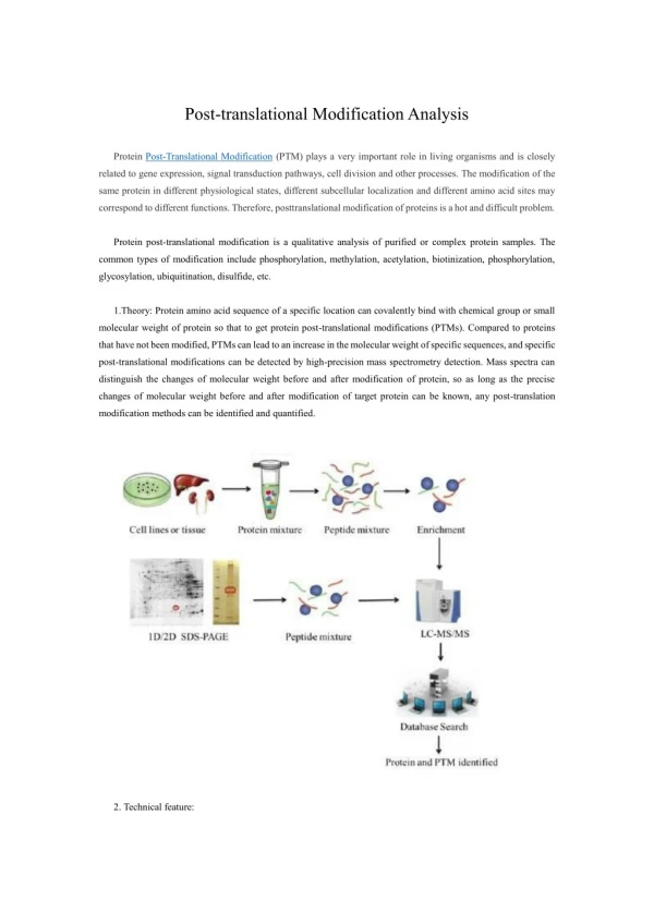
Identification of Post-translational Modifications For Sample Prep. Complexity of the Proteome. Protein processing and modification comprise an important third dimension of information, beyond those of DNA sequence and protein sequence.

E N D
Identification of Post-translational Modifications For Sample Prep
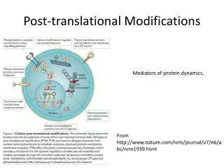
Complexity of the Proteome • Protein processing and modification comprise an important third dimension of information, beyond those of DNA sequence and protein sequence. • Many things can change cell components and their PTM’s: • cell cycle • environmental conditions • developmental stage • metabolic state. • Proteomic approaches don’t just identify proteins but also find their post-translational modifications are needed!
Post-translational Modification • What purpose ? -targeting (eg. some lipoproteins) -stability (eg. secreted glycoproteins ) - function (eg. surface glycoproteins) - control of activity (eg. clotting factors, caspases) • How can we study it ?
Definitions of the components: 1. Post-translational modification (PTM): Chemical modifications at certain amino acid residues - after the protein is synthesized by translation - are known as post-translational modifications. These are essential for normal functioning of the protein. Some of the most commonly observed PTMs include: a) Phosphorylation: The process by which a phosphate group is attached to certain amino acid side chains in the protein, most commonly serine, threonine and tyrosine. b) Glycosylation: The attachment of sugar moieties to nitrogen or oxygen atoms present in the side chains of amino acids like asparagine, serine or threonine. c) Acylation: The process by which an acyl group is linked to the side chain of amino acids like asparagine, glutamine or lysine. d) Alkylation: Addition of alkyl groups, most commonly a methyl group to amino acids such as lysine or arginine. Other longer chain alkyl groups may also be attached in some cases. e) Hydroxylation: This PTM is most often found on proline and lysine residues which make up the collagen tissue. It enables crosslinking and therefore strengthening of the muscle fibres.
Definitions of the components 2. Protein translation: The process by which the mRNA template is read by ribosomes to synthesize the corresponding protein molecule on the basis of the three letter codons, which code for specific amino acids. 3. Cytosol: A cellular compartment that serves as the site for protein synthesis. 4. Signal sequence: A sequence that helps in directing the newly synthesized polypeptide chain to its appropriate intracellular organelle. This sequence is most often cleaved following protein folding and PTM. 5. Endoplasmic reticulum: A membrane-bound cellular organelle that acts as a site for post-translational modification of the newly synthesized polypeptide chains. 6. Cleaved protein: The protein product obtained after removal of certain amino acid sequences such as N- or C-terminal sequences, signal sequence etc.
Proteomic analysis of PTMs Mann and Jensen, Nature Biotech. 21, 255 (2003)
Adduct formation – expect the unexpected …around 290 different adducts Statistics: Adducts in NIST12 MS/MS DB (80,000 spectra) Most common adducts for LC-MS ([M+H]+ [M+Na]+ [M+NH4]+ [M+acetate]+)
Different types of PTMs & their modification sites Ser, Thr, Tyr Pro, Lys Asn, Ser, Thr Lys, Arg Asn, Gln, Lys
Process of post-translational modification Translated Protein mRNA Ribosome Cytosol Endoplasmic reticulum (ER) Protein folding & PTMs Removal of certain N- and C-terminal residues Cleaved protein Protease Glc Glc P P CH3 CH3 Source: Modified from Biochemistry by A.L.Lehninger, 4th edition (ebook)
Increased complexity of proteome due to PTMs Gene sequence Expected protein structure Actual protein structure A C C A G C G U G A A A G A G G C C C C G C U C C U U P Glc Asn Asn Ala Ala Gly Gly His His Val Val Ala Ala Arg Arg CH3 Leu Leu Thr Thr
Phosphorylation reactions COO- COO- Kinase H R R R C OH C PO43- H O ATP ADP Amino acid residue Phosphorylated residue NH3+ NH3+ Ser CH2 Thr CH CH3 Tyr CH2 Source: Modified from Biochemistry by A.L.Lehninger, 4th edition (ebook)
Glycosylation reactions N-linked Glycosylation Sugar residues COO- COO- Glycosyl transferase R R C C H CONH2 CON OH O H NH3+ NH3+ Asn N-linked amino acid CH2 CH2 O-linked Glycosylation Glycosyl transferase COO- COO- H H C C Ser/Thr O-linked amino acid NH3+ NH3+ Source: Modified from Biochemistry by A.L.Lehninger, 4th edition (ebook)
Definitions of the components:Gel-based detection techniques for PTMs 1. Pro-Q-diamond: This fluorescent dye detects modified proteins that have been phosphorylated at serine, threonine or tyrosine residues. They are used with electrophoretic techniques and offer sensitivity down to few ng levels, depending upon the format in which they are used. This dye can also be combined with other staining procedures thereby allowing more than one detection protocol on a single gel. a) Gel staining: The process by which the protein bands on an electrophoresis gel are stained by suitable dyes for visualization. b) Gel scanning: The visualization of the stained protein bands on an electrophoresis gel by exciting it at a suitable maximum wavelength such that the dye absorbs the light and emits its own characteristic light at another emission wavelength. 2. Immunoblotting: This process, also known as Western blotting, is a commonly used analytical technique for detection of specific proteins in a given mixture by means of specific antibodies to the given target protein. a) Electrophoresis: Electrophoresis is a gel-based analytical technique that is used for separation and visualization of biomolecules like DNA, RNA and proteins based on their fragment lengths or charge-to-mass ratios using an electric field. The protein mixture is first separated by means of a suitable electrophoresis technique such as SDS-PAGE or Two-dimensional Electrophoresis.
Definitions of the components:Gel-based detection techniques for PTMs b) Blotting: The process by which the proteins separated on the electrophoresis gel are transferred on to another surface such as nitrocellulose by placing them in contact with each other. c) Nitrocellulose sheet: A membrane or sheet made of nitrocellulose onto which the protein bands separated by electrophoresis are transferred for further probing and analysis. d) Specific probe antibodies: Antibodies that are specific to a particular protein modification can be used as probes to detect those proteins containing that particular PTM. Protein phosphorylation is commonly detected using anti-phosphoserine, phosphothreonine or phosphotyrosine antibodies. Recently, specific motif antibodies have also been developed which detect a particular sequence of motif of the protein that contains a PTM. e) Labeled secondary Abs: Antibodies labeled with a suitable fluorescent dye molecule are used to detect the interaction between the modified protein and its antibody by binding to another domain of the probe antibody.
Pro-Q-diamond staining Completed 2-DE gel Dye stains the phosphorylated protein bands only. Tubing connected & outlet opened Protein bands get fixed on gel and minimize diffusion. Excess dye removed Tray with fixing solution (methanol + acetic acid) Pro-Q-diamond stain Washing solution (methanol + acetic acid)
Gel scanning Gel scanner Gel removed from scanner Stained gel Emission maxima – 580 nm Decreasing molecular weight Phosphoprotein image Decreasing pH
Dual staining with SYPRO-Ruby Red Tubing connected & outlet opened Dye stains all protein bands. Excess dye removed SYPRO-Ruby red staining solution Washing solution (methanol + acetic acid)
Gel scanning Gel scanner Phosphoprotein image Stained gel Emission maxima – 610 nm Total protein image Decreasing molecular weight Fluorescence Fluorescence A comparative profile between total protein image and phosphoprotein image enables detection of phosphorylated proteins. Decreasing pH Phosphoprotein image Total protein image by SYPRO-Ruby Red
Immunoblotting Proteins focused on IPG strip 2-D Electrophoresis SDS-PAGE Sample loading - Cathode Direction of migration Protein mixture Direction of migration Anode + Acrylamide gel Buffer Completed stained gels
Immunoblotting(this one for phosphorylated tyrosines!) Proteins phosphorylated at Tyr residues Specific phospho-tyrosine antibodies added Completed gels Nitrocellulose sheet or PVDF Detection using labeled secondary antibodies Blotting Proteins phosphorylated at Tyr residues
Phospho – ProteomicsWestern 2D gel , Ab specific to phospho-tyrosine
Phosphorylation and Mass Spec • Analysis of the entire complement of phosphorylated proteins in cells: “phosphoproteome” • Qualitative and quantitative information regarding protein phosphorylation important in many cellular processes • signal transduction, gene regulation, cell cycle, apoptosis • Most common sites of phosphorylation: Ser, Thr, Tyr • MS can be used to detect and map locations for phosphorylation • MW increase from addition of phosphate group • treatment with phosphatase allows determination of number of phosphate groups • digestion and tandem MS allows for determination of phosphorylation sites
Enrichment strategies to analyze phosphoproteins/peptides • Chemical derivatization • Introduce affinity tag to enrich for phosphorylated molecules • e.g., biotin binding to immobilized avidin/streptavidin
Enrichment strategies to analyze phosphoproteins/peptides • Oda et al., Nature Biotech. 2001, 19, 379 for analysis of pS and pT • Remove Cys-reactivity by oxidation with performic acid • Base hydrolysis induce ß-elimination of phosphate from pS/pT • Addition of ethanedithiol allows coupling to biotin • Avidin affinity chromatography to purify phosphoproteins AND MORE~!
Enrichment strategies to analyze phosphoproteins/peptides • Phosphospecific antibodies • Anti-pY quite successful • Anti-pS and anti-pT not as successful, but may be used (M. Grønborg, T. Z. Kristiansen, A. Stensballe, J. S. Andersen, O. Ohara, M. Mann, O. N. Jensen, and A. Pandey, “Approach for Identification of Serine/Threonine- phosphorylated Proteins by Enrichment with Phospho-specific Antibodies.” Mol. Cell. Proteomics 2002, 1:517–527. • Immobilized metal affinity chromatography (IMAC) • Negatively charged phosphate groups bind to postively charged metal ions (e.g., Fe3+, Ga3+) immobilized to a chromatographic support • Limitation: non-specific binding to acidic side chains (D, E) • Derivatize all peptides by methyl esterification to reduce non-specific binding by carboxylate groups. • Ficarro et al., Nature Biotech. (2002), 20, 301.
Phosphoprotein and Sypro Ruby Stains with Laser Imaging Beta-galactosidase Bovine serum albumin (BSA) Ovalbumin Beta-casein Avidin lysozyme PeppermintStickphosphoprotein molecular weight standards (LifeTechnologies) separated on a 13% SDS polyacrylamide gel. The gel was stained with Pro-Q Diamond phosphoprotein gel stain (blue) followed by SYPRO Ruby protein gel stain (red). The digital images were pseudocolored Phosphorylated BAPTA
Phosphoprotein Stain Visualization of total protein and phosphoproteins in a 2-D gel Proteins from a Jurkat T-cell lymphoma line cell lysate separated by 2-D gel electrophoresis and stained with Pro-Q Diamond phosphoprotein gel stain (blue) followed by SYPRO Ruby protein gel stain (red). After each dye staining, the gel was imaged and the resulting composite image was digitally pseudocolored and overlaid. T.H. Steinberg et al., Global quantitative phosphoprotein analysis using Multiplexed Proteomics technology, Proteomics 2003, 3, 1128-1144
Protein Glycosylation • The most important and complex form of PTM • Approx. 1% mammalian genes • Early view about carbohydrates (non-specific, static structures) has been challenged Ann. Rev. Biochem. 72(2003)643
Glycoprotein Gel Stain Detection of glycoproteins and total protein on an SDS-polyacrylamide gel using the Pro-Q Fuchsia Glycoprotein Gel Stain Kit. CandyCane glycoprotein molecular weight standards (LifeTechnologies) containing alternating glycosylated and nonglycosylated proteins electrophoresed through a 13% polyacrylamide gel. After separation, the gel was stained with SYPRO Ruby protein gel stain to detect all eight marker proteins (left). Subsequently, the gel was stained by the standard periodic acid–Schiff base (PAS) method in the Pro-Q Fuchsia Glycoprotein Gel Stain Kit to detect the glycoproteins alpha2-macroglobulin, glucose oxidase, alpha1-glycoprotein and avidin. Pro-Q™ Glycoprotein Stain (DDAO phosphate) Molecular Formula: C15H18Cl2N3O5P (MW 422.20)
Nitro-Tyrosine Modification • Oxidative modification of amino acid side chains: • methionine oxidation to the corresponding sulfone • S-nitrosationor S-nitrosoglutationylation of cysteine residues • Tyrosine modification to yield o,o’-dityrosine, 3-nitrotyrosine and 3-chlorotyrosine. • Tyrosine nitration is a well-established protein modification that occurs in disease states associated with oxidative stress and increased nitric oxide synthase activity. • The combination of 2D-PAGE, western blotting, IMMUNOASSAY and mass spectrometry has been the more typical strategy to identify 3-nitrotyrosine-modified proteins.
Nitro-Tyrosine Modification “Proteomic method identifies proteins nitrated in vivo during inflammatory challenge,” K. S. Aulak, M. Miyagi, L. Yan, K. A. West, D. Massillon, J. W. Crabb, and D. J. Stuehr, Proc. Natl. Acad. Sci. USA 2001; 98: 12056-12061. Anti-nitrotyrosine immunopositive proteins in lung of rats induced with LPS.
SERVICES at OSU Proteomics • Protein Growth, Induction and Expression, Protein purification • Subcloning into recombinant cell lines, Plasmid design • DIGE • Develop novel protein protocols, individualized for experiment • Selective subfractionation, Salt fractionation, Enrichment, Solubility screening, Inclusion body isolation • Western Blotting, Far Western Blotting, Immunoprecipitation and Co-immunoprecipitation, Protein-Protein interaction studies • Classic chromatography: • Affinity –Tag purification, ionic exchange, HIC reverse phase, SEC gel chromatography 100,300, Immobilized metal affinity chromatography (IMAC), Heparin affinity: Protein A/G affinity column, ENDOTOXIN removal • SDS-PAGE and DNA Electrophoresis, reduced and/or non-reduced • ProQ, LavaPurple, Sypro and other gel staining • Fluorescent and Bradford Protein Quantitation • Mass Spectrometry for protein identification Just ask! PTM identification!
THANKS FOR LISTENING! You can find us at… Mass Spec and Proteomics and Protein Expression and Purification Facility Biomedical Research Tower Room 250 460 West 12th Street Columbus, Ohio Lab: 614-247-8789 Arpad Somogyi, PhD – somogyi.16@osu.edu Cindy L. James, PhD – james.456@osu.edu