Download
1 / 65
650 likes | 692 Views
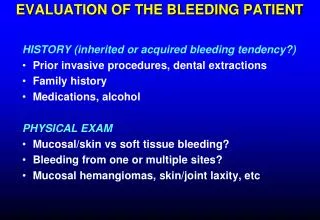
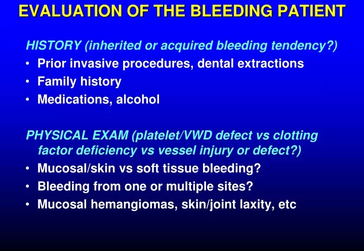
EVALUATION OF THE BLEEDING PATIENT. HISTORY (inherited or acquired bleeding tendency?) Prior invasive procedures, dental extractions Family history Medications, alcohol PHYSICAL EXAM (platelet/VWD defect vs clotting factor deficiency vs vessel injury or defect?)

E N D
EVALUATION OF THE BLEEDING PATIENT HISTORY (inherited or acquired bleeding tendency?) • Prior invasive procedures, dental extractions • Family history • Medications, alcohol PHYSICAL EXAM (platelet/VWD defect vs clotting factor deficiency vs vessel injury or defect?) • Mucosal/skin vs soft tissue bleeding? • Bleeding from one or multiple sites? • Mucosal hemangiomas, skin/joint laxity, etc
Laboratory tests to evaluate bleedingInitial screening • Platelet count • PT/INR • aPTT • Fibrinogen • Platelet function screen • Thromboelastography? Normal results → Major defect in primary hemostasis or clotting factor deficiency unlikely
Prothrombin time/INR Long PT/INR • Liver disease • Vitamin K deficiency • Warfarin or warfarin analogs (rat poison) • DOAC or high level of heparin • DIC • Inherited conditions rare • Won’t detect hemophilia, factor VIII inhibitor, heparin at therapeutic concentrations Magnitude of test abnormality usually proportional to clinical severity
aPTT Long aPTT with normal INR • Heparin (therapeutic or contaminant) or DOAC • Hemophilia A or B • von Willebrand disease (low VIII – but PTT may be normal) • Factor XI deficiency • Factor VIII inhibitor • Factor XII deficiency (does not cause bleeding) • Lupus anticoagulant (rarely causes bleeding) Magnitude of test abnormality not necessarily proportional to clinical severity
PFA-100 Platelet function screen • Replaces the bleeding time • Advantages • Ex vivo test (no skin incision) • Better standardized • Better sensitivity and specificity A patient with von Willebrand disease (VWF activity < 20%)
Sensitive to: Defective platelet adhesion in von Willebrand disease Platelet dysfunction due to drugs (variable sensitivity) Inherited platelet disorders Not useful in patients with moderate or severe Thrombocytopenia Platelet function screen
Conditions that may cause bleeding with normal or near-normal screening tests • Mild hemophilia (factor level 20-30% of normal) • Von Willebrand disease (mild variants) • Dysfibrinogenemia • Factor XIII deficiency (rare) • Fibrinolytic disorders (rare) • Vascular disorders (Ehlers-Danlos, amyloid, etc)
Laboratory tests to evaluate bleedingSecond round • Thrombin time (dysfibrinogenemia, heparin-like anticoagulant) • Von Willebrand activity • Alpha-2 antiplasmin (hyperfibrinolysis) • Factor XIII level • Platelet aggregation • Thromboelastography • Mixing study (if circulating inhibitor suspected)
WHY IS MY PATIENT BLEEDING? Blood vessel damaged/severed • History • Bleeding confined to area of injury • Coag tests normal or nearly so Potential intervention: sutures
Bleeding confined to an operative site is usually due to a severed vessel
WHY IS MY PATIENT BLEEDING? Defective primary hemostasis (platelets/VWF) • Skin/mucosal bleeding may be prominent • Petechiae (mainly with thrombocytopenia) • Bleeding soon after injury Lab results: abnormal platelet function screen, VWF activity, thromboelastography, platelet aggregometry Potential interventions: platelet transfusions, DDAVP, VWF concentrate
Platelet defects and vessel disorders: immediate bleeding from skin and mucosal surfaces, petechiae
WHY IS MY PATIENT BLEEDING? Defective fibrin formation (clotting factor deficiency or circulating anticoagulant) • Bleeding into tissues • Bleeding may be delayed after injury Lab results: Long PT/INR and/or PTT, low fibrinogen, abnormal TEG Potential interventions: FFP, cryoprecipitate, specific factor concentrate, prothrombin complex concentrate, vit K, rVIIa
Coagulation factor deficiency: delayed bleeding into soft tissues
WHY IS MY PATIENT BLEEDING? Hyperfibrinolysis • Generalized bleeding/oozing • Risk factors: liver disease, prostate cancer, leukemia, acute illness with DIC • Bleeding may be out of proportion to coag test abnormalities Lab results: low antiplasmin, abnormal TEG, low fibrinogen Potential intervention: antifibrinolytic drug
WHY IS MY PATIENT BLEEDING? Defective fibrin crosslinking (factor XIII deficiency) • Rare • Most cases hereditary & diagnosed in early childhood • Not detected by usual screening tests Lab result: low factor XIII Potential intervention: factor XIII concentrate
WHY IS MY PATIENT BLEEDING? Abnormal blood vessels • Examples: amyloidosis, Ehlers-Danlos syndrome, hereditary hemorrhagic telangiectasia • Coag tests usually normal • Exam findings often helpful
Hereditary hemorrhagic telangiectasia Researchgate.net
Ehlers-Danlos Syndrome Lifewitheds.com Morphopedics.wikdot.com
IMMUNE THROMBOCYTOPENIA • ITP • Drug-induced purpura • Heparin-induced thrombocytopenia • Post-transfusion purpura
ITP • Autoimmune platelet destruction • Mild to severe thrombocytopenia • Other blood counts, coag tests typically normal • A diagnosis of exclusion • Bleeding risk low to moderate in most cases • Hospitalization not always needed • High risk: “wet purpura”, active bleeding, elderly, history of major bleeding • Rarely begins in hospital • Few symptoms other than bruising & petechiae
ITP: initial treatment • Corticosteroids: prednisone 1 mg/kg or pulse high dose dexamethasone • IVIG (high dose) • Platelet transfusion sometimes effective • Splenectomy rarely necessary as initial treatment
Drug-induced thrombocytopenia • Thrombocytopenia may be severe (<5K) • Often begins in hospital, precipitous drop in platelet count • Bleeding risk moderate to high • Typically occurs within days of starting a drug
* * * * * * DRUGS MOST LIKELY TO CAUSE THROMBOCYTOPENIA Hematology 2009;153
Drug-induced thrombocytopeniamanagement • Stop any potentially offending drug • Resolution may take days to weeks • Steroids less effective than in ITP • IVIG worth trying if there is significant bleeding • Antifibrinolytics may help
Heparin-induced thrombocytopenia • Mild to moderate thrombocytopenia with onset 4-7 days after initial heparin exposure (UFH > LMWH) • Recent surgery, infection, inflammation increase risk • Bleeding risk low, thrombosis risk high • Heparin Ab test very sensitive – HIT can be ruled out if negative • Serotonin release assay predicts thrombotic risk • Rx: Stop heparin (any form), consider alternative anticoagulant (not warfarin or LMWH)
Post-transfusion purpura • Severe, precipitous drop in platelet count, usually a few days after blood product exposure (FFP, RBC most common) • Most patients are multigravid women • Prior exposure to platelet antigen during pregnancy, recall reaction after transfusion • Most affected patients lack the common platelet alloantigen HPA-1 (~ 2% of population) • Antibodies are directed against HPA-1 on transfused “passenger” platelets, patient’s own platelets are destroyed as “innocent bystanders” • Platelet transfusion may exacerbate the problem
Post-transfusion purpura • If suspected notify blood bank to arrange for appropriate testing and transfusion therapy • Test for HPA-1 antibodies in patient serum • Do NOT give platelet transfusion • If RBC transfusion needed give washed RBC • High dose IVIG • Steroids generally not helpful
DISSEMINATED INTRAVASCULAR COAGULATION • Rapid formation & lysis of intravascular fibrin • Consumption of clotting factors, platelets, inhibitors • Life-threatening underlying disease • Bleeding due to uncontrolled fibrinolysis, thrombocytopenia, clotting factor consumption and tissue injury from underlying disease • Tissue injury/necrosis due to microvascular occlusion, hypotension, cytokine-mediated endothelial damage • Most deaths due to underlying disease
PUPURA FULMINANS Pneumococcal sepsis in splenectomized patient NEJM 2001;344:1593
DIAGNOSIS OF DIC • Underlying disease capable of causing DIC? • Evidence of accelerated clotting factor and platelet consumption, and increased fibrinolysis? If both present DIC is likely
SCREENING FOR DIC • D-dimer (most sensitive) • PT/INR (PTT less helpful) • Fibrinogen • CBC/platelet count • Fibrin monomer (most specific)
TREAT UNDERLYING DISEASE! Clotting factor & inhibitor replacement if there is significant bleeding: Fresh frozen plasma (goal INR ≤ 1.8) Cryoprecipitate (goal fibrinogen ≥ 100) Platelets (goal platelet count 50-75K) Pharmacologic inhibitors (selected pts with major bleeding unresponsive to replacement) Heparin (low dose – eg, 500 U/hr) Antifibrinolytics (Amicar, tranexamic acid) TREATMENT OF DIC
TTP • Microangiopathic hemolytic anemia • Thrombocytopenia • Organ dysfunction (CNS, renal, other) due to small vessel occlusion • Untreated mortality rate >90% • With treatment mortality < 20% • 1-2 cases/yr @ UWHC
TTP • Caused by autoimmune destruction of ADAMTS-13 protease that modulates von Willebrand factor multimer size • Very large/sticky multimers clump platelets • Microthrombi damage RBC and block vessels
TTP • Thrombocytopenia (may be severe) • Intravascular hemolysis (high LDH, low haptoglobin, schistocytes in blood smear) • INR, PTT, fibrinogen usually normal • Organ dysfunction • Neurologic symptoms • Renal dysfunction (hematuria, proteinuria) • Cardiac (arrhythmia) • ADAMTS-13 activity low (usually <10%)
TTP - DDX • Occult cancer • Pregnancy complications (HELLP, etc) • Hemolytic-uremic syndrome (kidney main target organ) • Shiga toxin, GI prodrome • “Atypical” HUS: genetic component, complement-mediated, no GI prodrome • Vasculitis (SLE, scleroderma) • HIV infection • DIC • Drugs (calcineurin inhibitors, mitomycin C, interferon, clopidogrel, ticlopidine)
TTP • Urgent plasma exchange • Plasma infusion if PE not immediately available • Immunosuppression • Corticosteroids • Rituximab for patients with resistant, refractory or relapsed disease (or upfront?) • Do not transfuse platelets unless there is lifethreatening bleeding
HEMOPHILIA • Inherited deficiency of factor VIII (hemophilia A) or factor IX (hemophilia B) • Sex-linked inheritance; almost all patients male • Most bleeding into joints, muscles; mucosal and CNS bleeding uncommon • Severity inversely proportional to factor level: < 1%: severe, bleeding after minimal injury 1-5%: moderate, bleeding after mild injury > 5%: mild, bleeding after significant trauma or surgery (may not be diagnosed until adulthood)
HEMOPHILIATreatment of bleeding episodes • Unexplained pain in a hemophilia should be considered due to bleeding unless proven otherwise • External signs of bleeding may be absent • Treatment: factor replacement, pain control • Most adult patients self-administer factor • Test for inhibitor (antibody vs factor) if unexpectedly low response to factor replacement • Most inhibitors occur in children
TREATMENT OF BLEEDS IN HEMOPHILIA • Administer appropriate clotting factor concentrate • 20-40 U/kg for minor bleeds, repeat daily for 2-3 days • 40-50 U/kg for major bleeds, repeat daily for 4-7 days • 50 U/kg q 12 hours for life-threatening bleeds • 1 U/kg should increase plasma level of factor by about 2% • Initial dose higher with factor IX concentrate (greater volume of distribution, but longer half-life)
Emicuzumab – a new treatment for hemophilia A Uses: prevention of bleeding in patients with severe or moderate hemophilia A (given every 1-3 weeks) or to treat bleeding in patients with factor VIII inhibitors
TREATMENT OF HEMOPHILIACS WITH INHIBITORS • Call a hematologist • Recombinant factor VIIa • Emicuzumab (hemophilia A only) • High dose factor VIII (if low titer inhibitor) • Induce tolerance with daily factor infusion ± immunosuppression
ACQUIRED FACTOR VIII DEFICIENCY • Autoantibody to factor VIII • Most patients elderly • Extensive soft tissue or mucosal bleeding (different bleeding pattern than inherited hemophilia) • Laboratory: prolonged aPTT with normal INR, platelets • aPTT not corrected by mixing with normal plasma • Factor VIII activity typically < 10% • Bleeding risk not proportional to factor level • Treatment: rVIIa, immunosuppression • Call the hematologist now!
VON WILLEBRAND DISEASE • Quantitative or qualitative deficiency of von Willebrand factor (activity level 0-30%) • Defective platelet adhesion • Low factor VIII (not usually clinically significant) • Dominant inheritance (often positive family hx) • Mucosal bleeding, bruising – usually mild to moderate, rarely emergent except after surgery or trauma • Severe disease may have hemophilia-like symptoms due to low factor VIII
VON WILLEBRAND DISEASE • Diagnosis: von Willebrand factor activity most sensitive test • Most patients have abnormal platelet function screen • PTT may be long if factor VIII low enough • Levels <30% cause pathologic bleeding • Treatment: • DDAVP appropriate for most patients but need to demonstrate efficacy • If DDAVP known to be ineffective or patient has not had trial dose, use VWF-containing concentrate (Humate-P or equivalent); this is most appropriate therapy for VWD patient with major bleeding