Download
1 / 16
300 likes | 550 Views
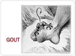
Gout. What are the risk factors for gout?. Hyperuricemia Male sex Older age Obesity Diet high in animal sources of purines (red meat, shellfish) Alcohol and high-fructose corn syrup-sweetened drinks Medications (thiazide or loop diuretics, cyclosporine) Renal insufficiency

E N D
What are the risk factors for gout? • Hyperuricemia • Male sex • Older age • Obesity • Diet high in animal sources of purines (red meat, shellfish) • Alcohol and high-fructose corn syrup-sweetened drinks • Medications (thiazide or loop diuretics, cyclosporine) • Renal insufficiency • Organ transplantation • Genetic risk factors
What comorbid diseases are associated with gout? • Renal insufficiency • Psoriasis • Hypertension • Diabetes • Hyperlipidemia • Metabolic syndrome • Cardiovascular disease
Are there effective strategies for prevention? • Dietary changes and weight loss • May lower serum urate levels • Therapy not indicated for asymptomatic hyperuricemia • Not proven to have adverse consequences • Long-term ULT may carry long-term risks • Treatment guidelines may change if there are sufficient evidence to show that hyperuricemia confers increased renal or cardiovascular disease risk
CLINICAL BOTTOM LINE: Prevention and Screening... • Risk factors • Hyperuricemia • Age, sex, obesity, renal insufficiency, diuretic use, diet • Genetic variants may increase risk • Common comorbidities • Diabetes, CVD, renal impairment, hypertension, metabolic syndrome, hyperlipidemia • Therapy not recommended for asymptomatic hyperuricemia • Lifestyle modifications appropriate in patients with only 1 gout attack and no other indications for ULT
What symptoms and physical examination findings suggest gout? • Acute onset joint pain at night • Swollen, erythematous, warm, exquisitely painful joint • Maximum pain within 24 h and resolves within 2 weeks • First Metatarsophalangeal joint most commonly involved • MSU crystals tend to form in previously diseased joints • With longer-disease duration and unabated hyperuricemia, persistent inflammation may occur • Urate deposition may be evident as subcutaneous nodules • Imaging may reveal tophaceous deposits
What tests can diagnose gout? • Examination of synovial fluid or tophus aspirate • Polarized microscopy, cell count, culture • MSU crystals in synovial fluid or tophus aspiration required to establish diagnosis • Other useful tests in diagnosing gout • Serum urate level • CBC with differential (if considering septic arthritis) • Radiography (to rule out other causes or to look for gouty erosions when symptoms are long-standing) • US or DECT imaging (to identify findings specific for gout)
What is the value of imaging? • Plain radiography • Show gout-related bone erosion, tophi • Show conditions coexisting with or confused for gout • Ultrasonography • Facilitate joint aspiration • Identify articular urate deposition, tophi, inflammation • DECT (not yet used in practice) • Differentiate calcium from urate • MRI (not routinely used in practice) • Show joint inflammation, damage, tophi—but cannot necessarily distinguish gout from CPP arthritis
What are the differential diagnoses? • Calcium pyrophosphate deposition • Septic arthritis • Cellulitis • Rheumatoid arthritis • Osteoarthritis • Psoriatic arthritis • Sarcoidosis
What classification criteria are used for gout in research studies? • MSU in synovial fluid or tophus aspiration is sufficient for classification as gout • ACR/EULAR criteria encompass following parameters: • Pattern of joint involvement during symptomatic episodes • Characteristics of symptomatic episodes • Time course of symptomatic episodes • Clinical evidence of tophus • Highest level of serum urate ever recorded off-treatment • MSU results of synovial fluid analysis • Imaging evidence of urate deposition • Imaging evidence of gout-related joint damage
CLINICAL BOTTOM LINE: Diagnosis... • MSU crystals in synovial fluid or tophus confirm diagnosis • Joint pain and hyperuricemia alone do not • Aspirate synovial fluid from joint or suspected tophus • Serum urate measurement is helpful but not diagnostic • Examine aspirated material under polarizing microscopy to differentiate gout from CPP-related arthritis • Examine synovial fluid cultures and clinical features to differentiate from septic arthritis • Radiography and ultrasonography: help identify other joint conditions and gout-specific features
When should clinicians consider hospitalizing a patient with gout? • Gout attacks are one of the most painful conditions • Hospitalization is warranted if: • Patient cannot care for self at home • Septic arthritis is a concern (to diagnose definitively and administer antibiotics promptly to prevent joint damage) • To monitor response to therapy, repeated synovial fluid analysis may be warranted for cell count and culture
What is the role of nonpharmacologic therapy in managing patients who already have gout? • Adjunct to long-term pharmacologic management • Most patients with gout require pharmacologic therapy • Lifestyle changes may help reduce serum urate levels • Reduce consumption of dietary contributors • Weight loss • Adequate hydration • Don’t blame patients for gout • Renal urate underexcretion, with genetic underpinnings, is the major contributor
What is the role of pharmacologic therapy? • Most patients require pharmacologic therapy • Urate-lowering therapy: cornerstone of management • Prophylaxis: when starting ULT to mitigate expected increased risk for attacks during initial phase • Anti-inflammatory therapy: for gout attacks • Indications for urate-lowering therapy • Frequent attacks (≥2 per year) • Tophus on clinical examination or imaging study • Chronic kidney disease stage ≥2 • Past urolithiasis (of any type)
When should clinicians consider consulting a specialist? • If a septic joint is suspected • To aid with joint aspiration • When gout is difficult to manage • First-line monotherapy insufficient • Contraindication or caution for gout attack management • Features may be related to other forms of arthritis • Patient is young, with possible inherited metabolic disease • Surgery is not indicated except when tophi pose an urgent function- or organ-threatening risk
CLINICAL BOTTOM LINE: Treatment... • Pharamcologic treatment • ULT if the patient has a clinical indication • Prophylaxis when initiating ULT • Anti-inflammatory therapy for gout attacks • Patient education • Causes of gout • Management of hyperuricemia • Adjunctive lifestyle modifications • Hospitalization warranted when gout-related pain and functional limitations cannot be controlled