Download
1 / 123
1.29k likes | 1.75k Views
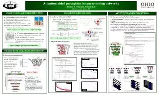
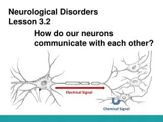
Section 2: Signal Transmission Between the Neurons. Neurotransmission. 1.Chemical synapse (Classical Synapse) Predominates in the vertebrate nervous system 2.Non-synaptic chemical transmission 3.Electrical synapse Via specialized gap junctions Does occur, but rare in vertebrate NS

E N D
Section 2: Signal Transmission Between the Neurons
Neurotransmission • 1.Chemical synapse (Classical Synapse) • Predominates in the vertebrate nervous system • 2.Non-synaptic chemical transmission • 3.Electrical synapse • Via specialized gap junctions • Does occur, but rare in vertebrate NS • Astrocytes can communicate via gap junctions
Chemical Synapse • Terminal bouton is separated from postsynaptic cell by synaptic cleft. • Vesicles fuse with axon membrane and NT released by exocytosis. • Amount of NTs released depends upon frequency of AP.
Non-synaptic chemical transmission The postganglionic neurons innervate the smooth muscles. No recognizable endplates or other postsynaptic specializations; The multiple branches are beaded with enlargements (varicosities) that are not covered by Schwann cells and contain synaptic vesicles; Fig. : Ending of postganglionic autonomic neurons on smooth muscle
Non-synaptic chemical transmission continued In noradrenergic neurons, the varicosities are about 5m, with up to 20,000 varicosities per neuron; Transmitter is apparently released at each varicosity, at many locations along each axon; One neuron innervate many effector cells. Fig. : Ending of postganglionic autonomic neurons on smooth muscle
Electrical Synapse • Impulses can be regenerated without interruption in adjacent cells. • Gap junctions: • Adjacent cells electrically coupled through a channel. • Each gap junction is composed of 12 connexin proteins. • Examples: • Smooth and cardiac muscles, brain, and glial cells.
Electrical Synapses • Electric current flow- communication takes place by flow of electric current directly from one neuron to the other • No synaptic cleft or vesicles cell membranes in direct contact • Communication not polarized- electric current can flow between cells in either direction
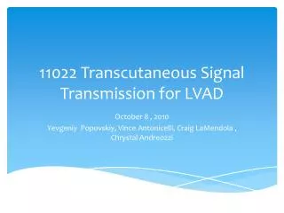
Chemical Synapse Electrical Synapse Purves, 2001
The chemical synapse is a specialized junction that transfers nerve impulse information from a pre synaptic membrane to a postsynaptic membrane using neurotransmitters and enzymes
Synaptic connections • ~100,000,000,000 neurons in human brain • Each neuron contacts ~1000 cells • Forms ~10,000 connections/cell • How many synapses?
Neurotransmitter- communication via a chemical intermediary called a neurotransmitter, released from one neuron and influences another • Synaptic cleft- a small gap between the sending (presynaptic) and the receiving (postsynaptic) site Chemical Synapses
Synaptic vesicles- small spherical or oval organelles contain chemical transmitter used in transmission • Polarization- communication occurs in only one direction, from sending presynaptic site, to receiving postsynaptic site Chemical Synapses
1. Synaptic Transmission Model • Precursor transport • NT synthesis • Storage • Release • Activation • Termination ~diffusion, degradation, uptake, autoreceptors
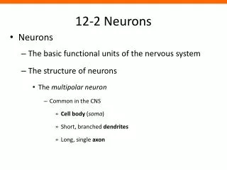
Postsynaptic Membrane Presynaptic Axon Terminal Terminal Button Dendritic Spine
(1) Precursor Transport
_ _ _ NT (2) Synthesis enzymes/cofactors
(3) Storage in vesicles
NT Vesicles Terminal Button Dendritic Spine Synapse
(4) Release Terminal Button Dendritic Spine Synapse Receptors
Terminal Button Dendritic Spine AP Synapse
Exocytosis Ca2+
Each vesicle contains one quanta of neurotransmitter (approximately 5000 molecules) –quanta release
(6.1) Termination by... Diffusion
(6.2) Termination by... Enzymatic degradation
(6.3) Termination by... Reuptake
(6.4) Termination by... Autoreceptors A
Autoreceptors • On presynaptic terminal • Binds NT • same as postsynaptic receptors • different receptor subtype • Decreases NT release & synthesis • Metabotropic receptors
Synaptic Transmission • AP travels down axon to bouton. • VG Ca2+ channels open. • Ca2+ enters bouton down concentration gradient. • Inward diffusion triggers rapid fusion of synaptic vesicles and release of NTs. • Ca2+ activates calmodulin, which activates protein kinase. • Protein kinase phosphorylates synapsins. • Synapsins aid in the fusion of synaptic vesicles.
Synaptic Transmission (continued) • NTs are released and diffuse across synaptic cleft. • NT (ligand) binds to specific receptor proteins in postsynaptic cell membrane. • Chemically-regulated gated ion channels open. • EPSP: depolarization. • IPSP: hyperpolarization. • Neurotransmitter inactivated to end transmission.
(1)Excitatory postsynaptic potential (EPSP) • An AP arriving in the presynaptic terminal cause the release of neurotransmitter; • The molecules bind and active receptor on the postsynaptic membrane;
(1)Excitatory postsynaptic potential (EPSP) • Opening transmitter-gated ions channels ( Na+) in postsynaptic- membrane; • Both an electrical and a concentration gradient driving Na+ into the cell; • The postsynaptic membrane will become depolarized(EPSP).
EPSP • No threshold. • Decreases resting membrane potential. • Closer to threshold. • Graded in magnitude. • Have no refractory period. • Can summate.
(2) Inhibitory postsynaptic potential (IPSP) • A impulse arriving in the presynaptic terminal causes the release of neurotransmitter; •The molecular bind and active receptors on the postsynaptic membrane open CI- or, sometimes K+ channels; • More CI- enters, K+ outer the cell, producing a hyperpolarization in the postsynaptic membrane.
(IPSPs): • No threshold. • Hyperpolarize postsynaptic membrane. • Increase membrane potential. • Can summate. • No refractory period.
3 Synaptic Inhibition • Presynaptic inhibition: • Amount of excitatory NT released is decreased by effects of second neuron, whose axon makes synapses with first neuron’s axon. • Postsynaptic inhibition
(1) Postsynaptic inhibition • Concept: effect of inhibitory synapses on the postsynaptic membrane. • Mechanism: IPSP, inhibitory interneuron • Types: Afferent collateral inhibition( reciprocal inhibition) Recurrent inhibition.
1) Reciprocal inhibition • Activity in the afferent fibers from the muscle spindles (stretch receptors) excites (EPSPs) directly the motor neurons supplying the muscle from which the impulses come. Postsynaptic inhibition At the same time, inhibits (ISPSs) those motor neurons supplying its antagonistic muscles.
1) Reciprocal inhibition The latter response is mediated by branches of the afferent fibers that end on the interneurons. Postsynaptic inhibition The interneurons, in turn, secrete the inhibitory transmitter (IPSP) at synapses on the proximal dendrites or cell bodies of the motor neurons that supply the antagonist.
Neurons may also inhibit themselves in a negative feedback fashion. Each spinal motor neuron regularly gives off a recurrent collateral that synapses with an inhibitory interneuron which terminates on the cell body of the spinal neuron and other spinal motor neurons. The inhibitory interneuron to secrete inhibitory mediator, slows and stops the discharge of the motor neuron. Postsynaptic inhibition 2) Recurrent inhibition
Concept: the inhibition occurs at the presynaptic terminals before the signal ever reaches the synapse. The basic structure: an axon-axon synapse (presynaptic synapse), A and B. Neuron A has no direct effect on neuron C, but it exert a Presynaptic effect on ability of B to Influence C. The presynatic effect May decrease the amount of neuro- transmitter released from B (Presynaptic inhibition) or increase it (presynaptic facilitation). (2) Presynaptic inhibition B A A A B C C
Presynaptic inhibition The mechanisms: • Activation of the presynaptic receptors increases CI- conductance, to decrease the size of the AP reaching the excitatory ending, reduces Ca2+ entry and consequently the amount of excitatory transmitter decreased. • Voltage-gated K+ channels are also opened, and the resulting K+ efflux also decreases the Ca2+ influx.
A B Presynaptic Inhibition Excitatory Synapse + • A active • B more likely to fire • Add a 3d neuron ~
A B - C Presynaptic Inhibition Excitatory Synapse + • Axon-axon synapse • C is inhibitory ~
A B - C Presynaptic Inhibition Excitatory Synapse + • C active • less NT from A when active • B less likely to fire ~