Download
1 / 22
310 likes | 1.41k Views
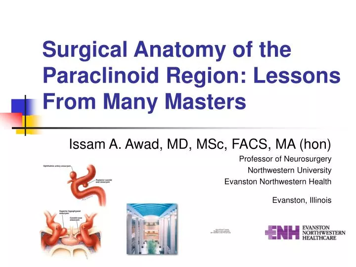
Surgical Anatomy of the Paraclinoid Region: Lessons From Many Masters . Issam A. Awad, MD, MSc, FACS, MA (hon) Professor of Neurosurgery Northwestern University Evanston Northwestern Health Evanston, Illinois. The Paraclinoid Region: Fundamentals for Every Surgeon.

E N D
Surgical Anatomy of the Paraclinoid Region: Lessons From Many Masters Issam A. Awad, MD, MSc, FACS, MA (hon) Professor of Neurosurgery Northwestern University Evanston Northwestern Health Evanston, Illinois
The Paraclinoid Region:Fundamentals for Every Surgeon • The anatomic facts: Rhoton’s Canon • Implications for paraclinoid aneurysms • Implications for surgical approach • Maximalist versus minimalist strategies • A personal philosophy
The Anatomic Facts: Rhoton’s Canon • Segments of the internal carotid artery (ICA) • Unique anatomic features of the C5-6 segments of the ICA • The oculomotor triangle • Relations to the optic nerve • Anatomy as the surgeon’s safeguard
The Anatomic Facts: Rhoton’s Canon • Segments of the ICA • Fisher • Berenstein and Lasjaunias • Bouthillier and van Loveren
The Anatomic Facts: Rhoton’s Canon • Unique anatomic features of the C5-6 segments of ICA
The Anatomic Facts: Rhoton’s Canon • Unique anatomic features of the C5-6 segments of ICA • Hemodynamic stresses • Imaging limitations • Dural relationships • Bony relationships • The subarachnoid space
The Anatomic Facts: Rhoton’s Canon • Unique anatomic features of the C5-6 segments of ICA • Hemodynamic stresses • Imaging limitations • Dural relationships • Bony relationships • The subarachnoid space
The Anatomic Facts: Rhoton’s Canon • Unique anatomic features of the C5-6 segments of ICA • Hemodynamic stresses • Imaging limitations • Dural relationships • Bony relationships • The subarachnoid space
Imaging The Paraclinoid Region Kobayashi: Cisternographic Guidance Gonzales, Zabramski and Spetzler: Optic Strut as Reference
The Anatomic Facts: Rhoton’s Canon • The oculomotor triangle • The interclinoid ligament • The tentorial edge (anterior petroclinoid ligament) • The posterior petroclinoid ligament • Relations to Cr. Ns. III, IV and VI
The Anatomic Facts: Rhoton’s Canon • The oculomotor triangle • The interclinoid ligament • The tentorial edge (anterior petroclinoid ligament) • The posterior petroclinoid ligament • Relations to Cr. Ns. III, IV and VI
The Anatomic Facts: Rhoton’s Canon • Relations to the optic nerve • The anterior clinoid process • The falciform ligament • The optic strut • The distal ring • The proximal ring
The Anatomic Facts: Rhoton’s Canon • Anatomy as the surgeon’s safeguard • Ease of approach • Vascular control • Maximize safety • Maximize exposure, maneuverability • Maximize effectiveness
Implications for Paraclinoid Aneurysms • The ophthalmic aneurysm • The superior hypophyseal aneurysm (extradural versus carotid cave) • The ventral paraclinoid aneurysm (transitional versus intradural)
Ophthalmic Aneurysm • Optic nerve canal decompression + clinoidectomy • Endovascular adjuncts • Proximal control • Suction decompression • Intraoperative angiography
Ophthalmic Aneurysm IO Angio IO Angio
Ventral Paraclinoid Aneurysm Clip Intradural Portion, Coil Extradural Portion
Maximalist vs. Minimalist Strategies • Adaptation of conventional approaches • Maximalist skull base approaches • Minimalist (keyhole, endoscopic assisted or controlled) • Focused strategies
A Personal Philosphy: Balancing What is “Safe”and What is “Feasible” • Proximal control • Intradural versus extradural consideration • Endovascular adjuncts • Endovascular treatments • Future challenges and opportunities -- surgical, endovascular