Download
1 / 20
210 likes | 431 Views
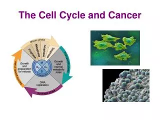
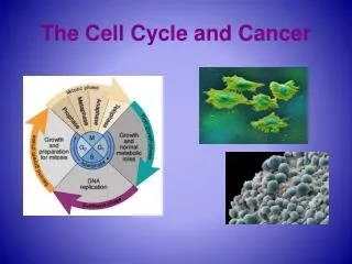
1. Cancer and the cell cycle checkpoints, reqmts to advance oncogenes tumor suppressor genes 2. 6 Traits of cancerous cells 3. Origins of cancerous cells. DNA. Mitotic Phase (M). DNA. DNA. DNA. Interphase. DNA. DNA. Cytokinesis. Mitosis. G 1 Cell growth.

E N D
1. Cancer and the cell cycle checkpoints, reqmts to advance oncogenes tumor suppressor genes 2. 6 Traits of cancerous cells 3. Origins of cancerous cells
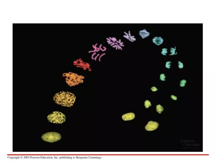
DNA Mitotic Phase (M) DNA DNA DNA Interphase DNA DNA Cytokinesis Mitosis G1 Cell growth G2 Cell growth preparation for division Interphase S DNA replication DNA DNA DNA Interphase
Proteins within the cell control the cell cycle • Signals affecting critical checkpoints determine whether the cell will divide (cyclins, kinases) G1 checkpoint Controlsystem M checkpoint Figure 8.9A G2 checkpoint
Anchorage, cell density, and chemical growth factors affect cell division • In laboratory cultures, normal cells divide only when attached to a surface = anchorage dependent
= density-dependent inhibition • Cells continue dividing until they touch one another Cells anchor to dish surface and divide. When cells have formed a complete single layer, they stop dividing (density-dependent inhibition). If some cells are scraped away, the remaining cells divide to fill the dish with a single layer and then stop (density-dependent inhibition). Figure 8.8A
Growth factors are proteins secreted by cells that stimulate other cells to divide After forming a single layer, cells have stopped dividing. Providing an additional supply of growth factors stimulates further cell division. Figure 8.8B
Growth factors bind to specific receptors on the plasma membrane to trigger cell division Growth factor Plasma membrane Relayproteins G1 checkpoint Receptor protein Signal transduction pathway Cell cyclecontrolsystem Figure 8.8B
Cancer cells have abnormal cell cycles • divide excessively and form tumors
Breast cancer cell - altered morphology Figure 8.10x1
Traits of cancer cells • 1. Independent of GROW signal from other cells often, oncogenes. Ex. ras • 2. Ignores STOP signal defective damage control, so problems not corrected. Often, tumor suppressor genes. Ex. p53
Traits of cancer cells, continued • 3. No cell suicide (apoptosis) If this occurs, treatments which damage dividing cells may not work. • 4. No limit to cell divisions telomeres rebuilt on ends of xsomes new treatment target: telomerase
Traits of cancer cells, continued • 5. Angiogenesis - formation of blood vessels • 6. Metastasis - ability to move to other tissues benign: do not move from tumor site malignant: invasive cells, can travel in blood and lymph system
Malignant tumors can invade other tissues and may kill the organism Lymphvessels Tumor Glandulartissue Metastasis 1 A tumor grows from a single cancer cell. 2 Cancer cells invade neighboring tissue. 3 Cancer cells spread through lymph and blood vessels to other parts of the body. Figure 8.10
How do normal cells become cancerous? Selection within tumor for “most cancerous” cells
What is the source of oncogenes? • Mutation of a normal gene = change in DNA sequence • UV light, Xrays, natural or synthetic chemicals • Virus (ex. HPV and cervical cancer)
Xsomal changes can be large or small Deletion Homologouschromosomes Duplication Inversion Reciprocaltranslocation Nonhomologouschromosomes Figure 8.23A, B
Xsomal translocation can activate an oncogene • A chromosomal translocation in the bone marrow • is associated with chronic myelogenous leukemia