Download
1 / 24
240 likes | 592 Views
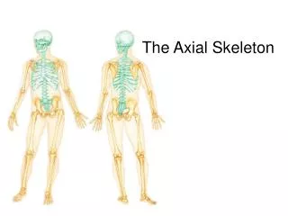
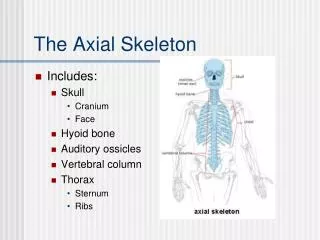
The Axial Skeleton. Access Human Biology. Task Label the diagram of the skeleton. Introduction. The skeleton is divided into two parts: The axial skeleton The appendicular skeleton

E N D
The Axial Skeleton Access Human Biology. Clare Hargreaves-Norris
TaskLabel the diagram of the skeleton Clare Hargreaves-Norris
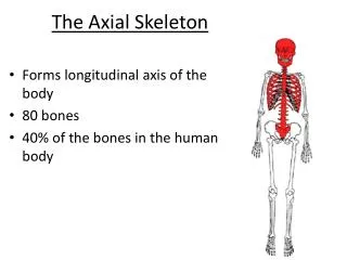
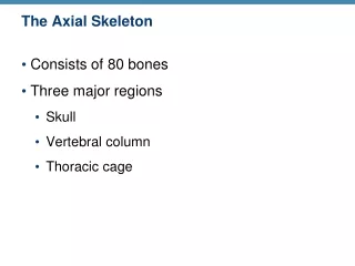
Introduction The skeleton is divided into two parts: • The axial skeleton • The appendicular skeleton The axial skeleton is made up of 80 bones comprising of the skull, rib cage and vertebral column. We will now look in detail at each section (see separate presentation for the skull). Clare Hargreaves-Norris
The Vertebral Column • The vertebral column is made up of 33 individual bones, though some are fused together. • In-between each vertebrae is a pad of fibrous tissue called a disc. • These discs act as shock absorbers to protect against gravity and injury. Clare Hargreaves-Norris
Functions Of The Vertebral Column • Protects the spinal cord – each vertebrae contains a hole in the centre through which the spinal cord runs. • Provides attachment for the ribs • Provides attachment for the muscles Clare Hargreaves-Norris
The first seven vertebrae are called the cervical vertebrae. At the top of the spinal column, these bones form a flexible framework for the neck and support the head. The first cervical vertebrae is called the atlas and the second is called the axis. The atlas shape allows the head to nod and the axis allows the head to shake. Cervical Vertebrae Clare Hargreaves-Norris
The next twelve vertebrae are called the thoracic vertebrae. These are found in the upper back. These bones move with the ribs to form the rear anchor of the ribcage. Thoracic vertebrae are larger than cervical vertebrae and increase in size from top to bottom. Thoracic Vertebrae Clare Hargreaves-Norris
The lumbar vertebrae are situated in the lower back. These five bones are the largest vertebrae in the spinal column. They support most of the body's weight and many of the back muscles attach to the lumbar vertebrae. Lumbar Vertebrae Clare Hargreaves-Norris
The sacrum is a triangular bone located just below the lumbar vertebrae. It consists of five sacral vertebrae in a child, which become fused into a single bone after age 26. The sacrum forms the rear wall of the pelvic girdle. The Sacrum Clare Hargreaves-Norris
The bottom of the spinal column is called the coccyx or tailbone. It consists of 4 bones that are fused together in an adult. Many muscles connect to the coccyx. The Coccyx Clare Hargreaves-Norris
When looked at from the side, the spine forms four curves. These curves are called the cervical, thoracic, lumbar, and pelvic curves. These curves allow human beings to stand upright and help to maintain balance. Curves Clare Hargreaves-Norris
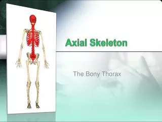
The ribs are thin, flat, curved bones that form a protective cage around the organs in the thorax. There are 24 bones arranged in 12 pairs. The ribs form a cage that encloses the upper body. The ribs protect the heart and lungs from injuries and shocks. The ribs also protect parts of the stomach, spleen, and kidneys. Ribs help you to breathe. Ribs Clare Hargreaves-Norris
The sternum is a flat, dagger shaped bone located in the middle of the chest. Along with the ribs, the sternum forms the rib cage. Sternum Clare Hargreaves-Norris
The Appendicular Skeleton ACCESS HE Human Biology Clare Hargreaves-Norris
Introduction The skeleton is divided into two parts: • The axial skeleton • The appendicular skeleton The appendicular skeleton is made up of 126 bones comprising of the arms, legs, pelvic girdle and shoulder girdle. Clare Hargreaves-Norris
There are two clavicles, more commonly known as the collar bones. These slender bones run from the shoulder to the breast bone, below the neck. Clavicle Clare Hargreaves-Norris
The two shoulder blades are situated in the upper back. Scapula Clare Hargreaves-Norris
The bone of the upper arm, one located in each arm. Humerus Clare Hargreaves-Norris
The radius runs from the elbow to the thumb side of the forearm. The ulna runs from the elbow to the little finger side of the forearm. Radius and Ulna Clare Hargreaves-Norris
The 16 carpal bones (8 in each) make up the bones of the wrist. There are 5 metacarpal bones in each hand. There are 28 phalanges that make up the fingers. Carpals, Metacarpals and Phalanges Clare Hargreaves-Norris
There are two bones that form the pelvic girdle. The pelvic girdle supports the weight of the body, protects and supports pelvic organs including: the bladder, the reproductive organs, and the developing fetus in a pregnant woman. The pelvic girdle differs between men and woman. In a man, the pelvis is larger and the iliac crests are closer together. In a woman, the pelvis is more delicate and the iliac crests are farther apart to allow for the birthing process. Pelvic bones Clare Hargreaves-Norris
There are two femur bones, one in each upper leg. The patella is the knee cap. Femur and Patella Clare Hargreaves-Norris
The tibia is one of the bones of the lower leg and runs medially to the big toe side of the leg. The fibula is the lateral bone of the lower leg. Tibia and Fibula Clare Hargreaves-Norris
There are 14 tarsal bones (7 in each ankle) that collectively form the ankle. There are 5 metatarsal bones in each foot. There are 28 phalanges that make up the toes. Tarsals, Metatarsals and Phalanges Clare Hargreaves-Norris