Download
1 / 63
710 likes | 1.12k Views
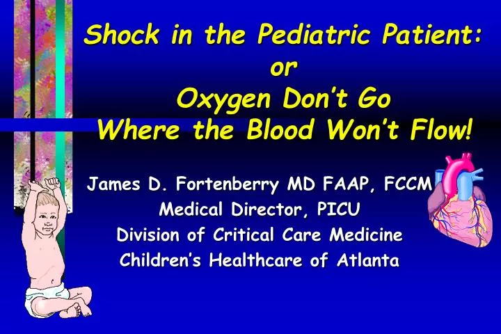
Shock in the Pediatric Patient: or Oxygen Don’t Go Where the Blood Won’t Flow!. James D. Fortenberry MD FAAP, FCCM Medical Director, PICU Division of Critical Care Medicine Children’s Healthcare of Atlanta. Objectives. Define shock and its different categories

E N D
Shock in the Pediatric Patient:orOxygen Don’t Go Where the Blood Won’t Flow! James D. Fortenberry MD FAAP, FCCM Medical Director, PICU Division of Critical Care Medicine Children’s Healthcare of Atlanta
Objectives • Define shock and its different categories • Review basic physiologic aspects of shock • Describe management of shock including: • oxygen supply and demand • fluid resuscitation • crystalloid vs. colloid controversy • vasopressor support
Definition of Shock • Uncontrolled blood or fluid loss • Blood pressure less than 5th percentile for age • Altered mental status, low urine output, poor capillary refill • None of the above
Definition of Shock An acute complex pathophysiologic state of circulatory dysfunction which results in a failure of the organism to deliver sufficient amounts of oxygen and other nutrients to satisfy the requirements of tissue beds
Definition of Shock • Inadequate tissue perfusion to meet tissue demands • Usually result of inadequate blood flow and/or oxygen delivery • Shock is not a blood pressure diagnosis!!
Characteristics of Shock • End organ dysfunction: • reduced urine output • altered mental status • poor peripheral perfusion • Metabolic dysfunction: • acidosis • altered metabolic demands
Essentials of Life • Gas exchange capability of lungs • Hemoglobin • Oxygen content • Cardiac output • Tissues to utilize substrate
Arterial Oxygen Content 100 mm Hg PaO2 100 mmHg Partial Pressure SaO2 97% Oxygen Saturation + Hgb 15 gm/100 mL Hemoglobin + O2 in plasma O2 bound to Hgb
Oxygen Delivery DO2=Cardiac Output x 1.34 (Hgb x SaO2) + Pa02 x 0.003 O2O2O2O2O2O2 Oxygen Express O2O2O2O2O2O2 Ca02
Cardiac Output The volume of blood ejected by the heart in one minute 4 - 8 liters / minute
Cardiac OutputC.O.=Heart Rate x Stroke Volume • Heart rate • Stroke volume: • Preload- volume of blood in ventricle • Afterload- resistance to contraction • Contractility- force applied
Cardiac OutputC.O.=Mean arterial pressure (MAP) - CVP/SVR • To improve CO: • MAP • CVP • SVR
Preload Afterload Contractility x Heart Rate Stroke Volume Cardiac Output O2 Content Resistance x x O2 Delivery Arterial Blood Pressure
Hypovolemic dehydration,burns, hemorrhage Distributive septic, anaphylactic, spinal Cardiogenic myocarditis,dysrhythmia Obstructive tamponade,pneumothorax Compensated organ perfusion is maintained Uncompensated Circulatory failure with end organ dysfunction Irreversible Irreparable loss of essential organs Classification of Shock
Mechanical Requirements for Adequate Tissue Perfusion • Fluid • Pump • Vessels • Flow
Hypovolemic Shock: Inadequate Fluid Volume (decreased preload)
Hypovolemic Shock:Causes • Fluid depletion • internal • external • Hemorrhage • internal • external
Cardiogenic Shock: Pump Malfunction (decreased contractility)
Cardiogenic Shock:Causes Electrical Failure • Mechanical Failure • Cardiomyopathy • metabolic • anatomic • hypoxia/ischemia
Distributive Shock Abnormal Vessel Tone (decreased afterload)
Distributive Shock Vasodilation Venous Pooling Decreased Preload Maldistribution of regional blood flow
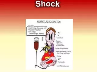
Distributive Shock: Causes • Sepsis • Anaphylaxis • Neurogenesis (spinal) • Drug intoxication (TCA, calcium, Channel blocker)
Septic Shock Decreased Pump Function Decreased Volume Abnormal Vessel Tone
Cardiac OutputC.O.=Heart Rate x Stroke Volume • Heart rate • Stroke volume: • Preload- volume of blood in ventricle • Afterload- resistance to contraction • Contractility- force applied
Clinical Assessment • Heart rate • Peripheral circulation • capillary refill • pulses • extremity temperature • Pulmonary • End organ perfusion • brain • kidney
Improving Stroke Volume:Therapy for Cardiovascular Support Preload Volume Inotropes Contractility Vasodilators Afterload
Septic Shock Early (“Warm”) Decreased peripheral vascular resistance Increased cardiac output Late (“Cold”) Increased peripheral vascular resistance Decreased cardiac output
Heart Rate and Perfusion Pressure (MAP-CVP) Parameters by Age
OBSTRUCTIVE SHOCK OBSTRUCTED FLOW
Obstructive Shock:Causes • Pericardial tamponade • Pulmonary embolism • Pulmonary hypertension
O2 content Cardiac output Blood pressure Goals of Resuscitation • Overall goal: • increase O2 delivery • decrease demand Treatment Sedation/analgesia
Principles of Management • A: Airway • patent upper airway • B: Breathing • adequate ventilation and oxygenation • C: Circulation • optimize • cardiac function • oxygenation
Act quickly,Think slowly. Greek Proverb
Airway Management • Patients in shock have: • O2 delivery • progressive respiratory fatigue/failure • energy shunted from vital organs • afterload
Airway Management • Early intubation provides: • O2 delivery and content • controlled ventilation which: • reduces metabolic demand • allows C.O. to vital organs
Therapy Vagolysis Heart Rate Chromotropy
Fluid Choices Colloid Crystalloid Less Filling Tastes Great !
CrystalloidsHypotonic Fluids (D5 1/4 NS) • No role in resuscitation • Maintenance fluids only
Fluids, Fluids, Fluids • Key to most resuscitative efforts • Give generously and reassess
CrystalloidsIsotonic Fluids • Intravascular volume expansion • Hauser: • crystalloids rapidly redistribute • Lethal animal model • NS = good resuscitative fluid • 4x blood volume to restore hemodynamics
CrystalloidsIsotonic Fluids • 2 trauma studies • crystalloids = colloids but: • 4x amount • longer time to resuscitation
CrystalloidsComplications • Under-resuscitation • renal failure • Over-resuscitation • pulmonary edema • peripheral edema
CrystalloidsSummary • Crystalloids less effective than equal volume of colloids • Preferred when 1o deficit is water and/or electrolytes • Good in initial resuscitation to restore extracellular volume • Hypertonic solutions however, may act as plasma volume expanders
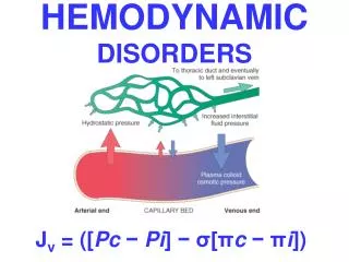
Oncotic pressure (tendency to pull unit) Hydrostatic pressure (tendency to drive unit) Fluid Transport Capillary
ColloidsAlbumin • Hepatic production • MW = 69,000 • 80% of COP • Serum t1/2: 18 hours endogenous 16 hoursexogenous
ColloidsHydroxyethyl Starch (Hespan) • Synthetic • Derived from corn starch • Average MW = 69,000 • Stable, nonantigenic • Used for volume expansion • Renal excretion • t 1/2 2-67 hours • 90% gone in 42 days