Download
1 / 22
260 likes | 1.07k Views
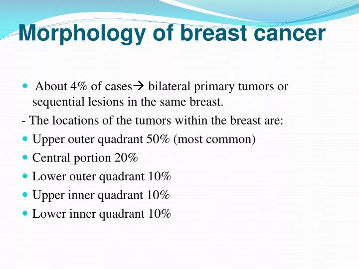
Morphology of breast cancer. About 4% of cases bilateral primary tumors or sequential lesions in the same breast. - The locations of the tumors within the breast are: Upper outer quadrant 50% (most common) Central portion 20% Lower outer quadrant 10% Upper inner quadrant 10%

E N D
Morphology of breast cancer • About 4% of cases bilateral primary tumors or sequential lesions in the same breast. - The locations of the tumors within the breast are: • Upper outer quadrant 50% (most common) • Central portion 20% • Lower outer quadrant 10% • Upper inner quadrant 10% • Lower inner quadrant 10%
Breast cancers are classified into: • Noninvasive (confined by a basement membrane and do not invade into stroma or lymphovascular channels), include: • Ductal carcinoma in situ (DCIS) • Lobular carcinoma in situ (LCIS) • Invasive (infiltrating) • Invasive ductal carcinoma – NOS (most common type) • Invasive lobular carcinoma • Medullary carcinoma • Colloid (mucinous) carcinoma • Tubular carcinoma • Other types
Ductal carcinoma in-situ DCIS • Nuclear appearance ranges from low nuclear grade to pleomorphic (high nuclear grade). • The comedo subtype: cells with high-grade nuclei distending ducts with extensive central necrosis. (The name derives from the toothpaste-like necrotic tissue). • Calcifications are frequently associated with DCIS • The detection of DCIS screening by mammography (calcifications). • The neoplastic cells express ER and PR. • The prognosis : excellent (97% long-term survival after simple mastectomy) • Current treatment strategies: surgery and radiation, tamoxifen • Significance: adjacent invasive CA; recurrence; become invasive if untreated
Paget disease of the nipple • is caused by the extension of DCIS up to the lactiferous ducts and into the contiguous skin of the nipple. The malignant cells disrupt the normal epidermal barrier, which allows extracellular fluid to be extruded onto the surface. • The clinical appearance unilateral crusting exudate over the nipple and areolar skin. • 50% of cases underlying invasive ca will be present. • Prognosis is based on the underlying carcinoma and is not worsened by the presence of Paget disease.
Lobular carcinoma in-situ LCIS • The cells fill and distend the lobules • rarely associated with calcifications. • 1/3 of women with LCIS eventually develop invasive carcinoma. • subsequent invasive carcinomas arise in either breast at significant frequency. (1/3 will be of lobular type (as compared with ∼10% of cancers in women who develop de novo lobular carcinoma), but most are of no special type. • LCIS is both a marker of increased risk of developing breast cancer in either breast and a direct precursor of some cancers. • Current treatment: close clinical and radiologic follow-up of both breasts or bilateral prophylactic mastectomy.
Invasive ductal carcinoma • Definition. A term used for all carcinomas that cannot be subclassified into one of the specialized types described later. • Also called Carcinomas "not otherwise specified" • 70% to 80% of all cancers fall into this group. • Precancerous lesion: usually associated with DCIS, but rarely LCIS • Clinical presentation: a mammographic density; a hard, palpable mass. Advanced cancers may cause dimpling of the skin, retraction of the nipple, or fixation to the chest wall. • Microscopic appearance: ranges from tumors with well-developed tubule formation and low-grade nuclei to tumors consisting of sheets of anaplastic cells. The tumor margins are usually irregular. Invasion of lymphovascular spaces or along nerves may be seen. • Receptor profile: 2/3 express ER or PR; 1/3 overexpresses HER2/NEU.
Invasive lobular carcinoma • Definition . Invasive cancer that consists of cells morphologically identical to the cells of LCIS. • comprise fewer than 20% of all breast carcinomas. • Precancerous lesion. 2/3of the cases adjacent LCIS. • Lobular carcinomas are also more frequently multicentric and bilateral (10% to 20%). • Microscopic picture. The cells invade individually into stroma and are often aligned in strands or chains. • Clinical presentation. Most present as palpable masses or mammographic densities, a significant subgroup may have a diffusely invasive pattern without a desmoplastic response and may be clinically occult. • Lobular carcinomas, more frequently than ductal carcinomas, metastasize to cerebrospinal fluid, serosal surfaces, gastrointestinal tract, ovary and uterus, and bone marrow. • Almost all of these carcinomas express hormone receptors, but HER2/NEU overexpression is very rare or absent.
Medullary carcinoma • A rare subtype of carcinoma <1% of cases. • Microscopically, These cancers consist of sheets of large anaplastic cells with pushing, well-circumscribed borders. There is a pronounced lymphoplasmacytic infiltrate. • Precancerouslesion:DCIS is usually absent or minimal. • Medullary carcinomas, occur with increased frequency in women with BRCA1 mutations, although most women with medullary carcinoma are not carriers. • Receptorprofile. These carcinomas uniformly lack hormone receptors and do not overexpress HER2/NEU.
Colloid (mucinous) carcinoma • is also a rare subtype. • Clinicalpicture. Like medullary carcinomas, they often present as well-circumscribed masses and can be mistaken for fibroadenomas. • Microscopicpicture. The tumor cells produce abundant quantities of extracellular mucin that dissects into the surrounding stroma. Grossly the tumors are usually soft and gelatinous. • Most express hormone receptors (ER,PR), and rare examples may overexpress HER2/NEU.
Tubular carcinomas • account for 10% of invasive carcinomas • Clinicalpresentation. They usually present as irregular mammographic densities. • Microscopically, the carcinomas consist of well-formed tubules with low-grade nuclei. • Lymph node metastases are rare, and prognosis is excellent. • Virtually all tubular carcinomas express hormone receptors, but overexpression of HER2/NEU is highly unusual.
Features Common to All Invasive Cancers • These include: • Fixation: tendency to become adherent to the pectoral muscles or deep fascia of the chest wall • retraction or dimpling of the skin or nipple: adherence to the overlying skin important sign, because it may be the first indication of a lesion, observed by the woman herself during self-examination. • peaud'orange (orange peel): Involvement of the lymphatic pathways cause localized lymphedema, the skin becomes thickened around exaggerated hair follicles
Spread of Breast Cancer • through lymphatic and hematogenous channels. - Outer quadrant and centrallyaxillary nodes. - inner quadrants tumors internal mammary lymph nodes • Favored locations are the lungs, skeleton, liver, and adrenals and (less commonly) the brain, spleen, and pituitary. • Metastases may appear many years after apparent therapeutic control of the primary lesion, sometimes 15 years later. • SCREENING • mammographic screening: - Can detect invasive carcinoma <1 cm (only 15% of these have nodal metastases). - DCIS is detected before the development of invasive carcinoma - The current controversy over the best time to begin mammographic screening ; usually at 35- 40 yr. • Magnetic resonance imaging MRI
Prognosis- depends on: 1- The size of primary carcinoma. 2- Lymph node involvement and the number of lymph nodes involved by metastases. (no axillary node involvement= 5-yr survival rate 90%). The survival rate decreases with each involved lymph node 3- Distantmetastases. 4- The grade of the carcinoma 5- The histologic type of carcinoma(tubular, medullary, and mucinous) have a better prognosis than "ductal carcinomas- NOS"). 6- The presence or absence of estrogen or progesterone receptors. The presence of hormone receptors confers a better prognosis 7- The proliferative rate of the cancer. 8- Aneuploidy.worse prognosis. 9- Overexpression of HER2/NEU: associated with a poorer prognosis. • the importance of evaluating HER2/NEU is to predict response to a monoclonal antibody ("Herceptin") against the gene product.
Male breast pathology Gynecomastia • Enlargement of the male breast • due to absolute or relative estrogen excesses. • Morphology: similar to those of intraductal hyperplasia. • usually in both breasts but occasionally in only one. • According to cause, divided into: 1- pathologic gynecomastia: cirrhosis of the liver; Klinefelter syndrome; estrogen-secreting tumors; estrogen therapy; digitalis therapy. 2- Physiologic gynecomastia: puberty and extreme old age.
Carcinoma of the male breast • frequency ratio to breast cancer in the female of 1: 125. • advanced age. • Both morphologically and biologically, resemble invasive carcinomas in the female. • Because of the scant amount of breast substance in the male, the tumor rapidly infiltrates the overlying skin and underlying thoracic wall. • Unfortunately, almost 1/2 have spread to regional lymph nodes and more distant sites by the time they are discovered.