Download
1 / 18
200 likes | 408 Views
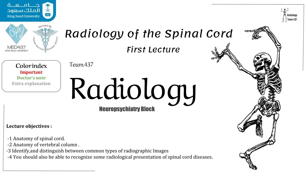
Radiology of the Spinal Cord. First Lecture. Team 437 Radiology. Color index Important Doctor’s note Extra explanation. Neuropsychiatry Block. Lecture objectives: 1- Anatomy of spinal cord . 2- Anatomy of vertebral column.

E N D
Radiology of the Spinal Cord First Lecture Team 437Radiology Color indexImportant Doctor’s noteExtra explanation Neuropsychiatry Block Lecture objectives: 1- Anatomy of spinal cord . 2- Anatomy of vertebral column. 3-Identify,and distinguish between common types of radiographic Images 4- You should also be able to recognize some radiological presentation of spinal cord diseases.
X-RAYS: Characteristics: -Often the first diagnostic imaging test -Small dose of radiation to visualize the bony parts -X-ray can detect: • Spinal alignment and curvature • Spinal instability-with flexion and extension views • Congenital (birth) defects of spinal column • Fracture caused by trauma • Moderate osteoporosis (loss of calcium from the bone) • Infections Not always • Tumors Not always -X-ray image of a person who suffers from Scoliosis
Open mouth view Lateral view Anteroposterior (frontal view) C1 C1 C2
Is this film an adequate(acceptable) lateral film? It’s not an adequate film because the 7th cervical vertebra is not seen in the image.
Computerized Tomography (CT SCAN): -Uses radiation -Obtain 2-D images (can be processed to 3-D images) -Entire spine can be imaged within a few minutes -Detailed information regarding bony structures -Limited information about spinal cord & soft tissues
Magnetic Resonance Imaging (MRI): T1 Weighted -Gold standard of imaging for spinal cord disorders -No radiation -Can identify abnormalities of bone, soft tissues and spinal cord -Claustrophobic(fear of small places) patients, uncooperative and children may need sedation or general anesthesia -Contraindications include implanted devices e.g. cardiac pacemakers and electromagnetic devices T2 Weighted *CSF *Cerebrospinal fluid appears white when taken by the T2 MRI method, while the T1 method makes the CSF appear darker.
Abnormalities Of Spinal Cord: Congenital Trauma Tumors demyelination Trauma Usually the first series of images to be ordered by the physician Plain Radiographs(x-rays). If fractures, or other bony defects are suspected CT imagescan provide very detailed information. When soft tissue injury is suspected MRI is usually the imaging technology of choice. . Important Accesses for parallel lines (imaginary lines): 1- Anterior vertebral line 2- Posterior vertebral line 3- Spinolaminar line 4- Posterior spinous line Any malalignment should be considered as an evidence of injury.
Mechanism Of Injury For trauma there is several mechanism of injury Here is some examples: Hyperextension: a sudden backward acceleration of the skull creating extreme extension of cervical spines ,a common example is like hanging, or when the head hits the dashboard of the car in a car accidents. Hyperflexion: excessive flexion of the neck like when diving in shallow water ,could cause paraplegia Compression Fracture Type of imaging in All pictures listed below is X-ray When you get a caudally directed force on your head (like when something heavy falls on yours head). Lateral mass splitting, displacement more than 2ml Normal open mouth view
Type of imaging in All pictures listed below is X-ray Hangman's Fracture (result of hyperextension) A fracture which involves the pars interarticularis of C2 on both sides, and a dislocation of C2 , its a result of hyperextension and distraction Normal lateral view Hyperflexion * *The distance between spinal processes is wide Normal lateral view A tear drop fracture in the anterior marginof the vertebral body
Congenital defects Spina bifida Syringomyelia -Its when a pregnant mother has deficiency of folic acid then the babies neural tube gets defected. for more information about the disease . A B C b) meningocele, MRI T1 WI c) meningomyelocele MRI T2 WI. a)Spina bifida occulta., CT scan -it is the development of a fluid-filled cyst within the spinal cord.
demyelination Multiple Sclerosis Multiple sclerosis (MS) is a relatively commonly acquired chronic relapsing demyelinating disease involving the CNS. Characteristically disseminated not only in space but also in time. Transverse Myelitis -Spinal cord is swollen. inflamed cord of uncertain cause • Viral infections • Immune reactions • Idiopathic Myelopathy progressing over hours to weeks.
Tumors Classification of TUMORS -Intramedullary lesions: its location is determined within the cord -Extramedullary lesions: May be related to nerve roots and may extend into the foramen (e.g. schwannomas and neurofibromas) or may have a broad dural attachment (e.g. meningiomas). Astrocytoma Ependymoma -Intramedullary tumor -Intramedullary tumor -There’s a chance of bleeding
MCQs: 1-Technology that can Obtain 2-D images (can be processed to 3-D images). A- X-RAY B-MRI C- CT SCAN D- ULTRASound 2-is a relatively common acquired chronic relapsingdemyelinating disease involving the CNS A-Multiple Sclerosis B-Ependymoma C-Astrocytoma D-Transverse Myelitis 3-inflamed cord of uncertain cause A-Multiple Sclerosis B-Ependymoma C-Astrocytoma D-Transverse Myelitis 4-When soft tissue injury is suspected, ___is usually the imaging Modality of choice. A- MRI B- XRAY C- CT SCAN D- ULTRASOUND Answers: 1- C 2- A 3- D 4- A
Thank you for checking our work Team Leaders: Faisal AlqusaiyerAljoharah Alshunaifi Team members: Afnan Almustafa Adel alzahraniRawan Alrehaili Abdullah alserganiAbeer Alabduljabbar Saad alhaddab Contact us on: @Radiology437 Radiology437@gmail.com