Download
1 / 1
10 likes | 212 Views
INTRAOPERATORY ASSESSMENT OF SENTINEL NODE IN BREAST CANCER PERFORMING ONE STEP NUCLEIC ACID AMPLIFICATION (OSNA) ASSAY ON THE WHOLE LYMPH NODE Sapino A., Macrì L, Lupo R, Marazzi A, Pecchioni C, De Rosa P and Castellano I

E N D
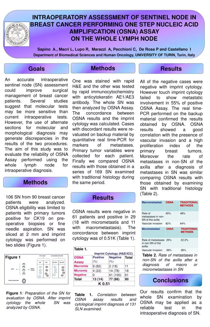
INTRAOPERATORY ASSESSMENT OF SENTINEL NODE IN BREAST CANCER PERFORMING ONE STEP NUCLEIC ACID AMPLIFICATION (OSNA) ASSAY ON THE WHOLE LYMPH NODE Sapino A., Macrì L, Lupo R, Marazzi A, Pecchioni C, De Rosa P and Castellano I Department of Biomedical Sciences and Human Oncology, UNIVERSITY OF TURIN, Turin, Italy Methods Goals Results An accurate intraoperative sentinel node (SN) assessment could improve surgical management of breast cancer patients. Several studies suggest that molecular tests may be more sensitive than current intraoperative tests. However, the use of alternate sections for molecular and morphological diagnosis may generate discrepancies in the results of the two procedures. The aim of this study was to evaluate the reliability of OSNA Assay performed using the whole lymph node for intraoperative diagnosis. One was stained with rapid H&E and the other was tested by rapid immunocytochemistry with anticytokeratin AE1/AE3 antibody. The whole SN was then analyzed by OSNA Assay. The concordance between OSNA results and the imprint cytology was calculated. Cases with discordant results were re-valuated on backup material by quantitative real time-PCR for markers of metastases. Primary tumor variables were collected for each patient. Finally we compared OSNA results with those obtained in a series of 169 SN examined with traditional histology during the same period. All of the negative cases were negative with imprint cytology. However touch imprint cytology failed to show metastatic involvement in 55% of positive OSNA Assay. The real time-PCR performed on the backup material confirmed the results obtained by OSNA. OSNA results showed a good correlation with the presence of vascular invasion and a high proliferation index of the primary breast tumors. Moreover the rate of metastases in non-SN of the axilla after a diagnosis of metastases in SN was similar comparing OSNA results with those obtained by examining SN with traditional histology (Table 2). Methods Figure 1 Results 106 SN from 90 breast cancer patients were analyzed. OSNA eligibility was limited to patients with primary tumors positive for CK19 on pre-operative biopsies or fine needle aspiration. SN was sliced at 2 mm and imprint cytology was performed on two slides (Figure 1). OSNA results were negative in 61 patients and positive in 29 (18 with micrometastis and 11 withmacrometastasis). The concordance between imprint cytology was of 0.51K (Table 1). Table 2. Rate of metastases in non-SN of the axilla after a diagnosis of macro or micrometastases in SN Conclusions Our results confirm that the whole SN examination by OSNA may be applied as a reliable test in the intraoperative diagnosis of SN. Figure 1: Preparation of the SN for evaluation by OSNA. After imprint cytology the whole SN was analyzed by OSNA. Table 1. Correlation between OSNA assay results and cytological imprint diagnosis of 131 SLN examined.