Download

1 / 25
250 likes | 474 Views
Male Reproductive System. January 30, 2008. Ductus deferens. Epididymis. Head. Body. Tail. From Netter. Slide 270. From J.M. Velkey. #270 Seminiferous tubules A. Spermatogonia B. Primary spermatocytes C. Spermatids D. Sertoli cells E. Leydig cells . A. D. C. E. C. B.

E N D
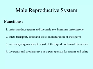
Male Reproductive System January 30, 2008
Ductus deferens Epididymis Head Body Tail From Netter
Slide 270 From J.M. Velkey
#270 Seminiferous tubulesA. Spermatogonia B. Primary spermatocytes C. Spermatids D. Sertoli cells E. Leydig cells A D C E C B E
UCSF slide 363 seminiferous tubules From J.M. Velkey
#275 Meiastinum testis and Epididymis Epididymis Efferent ducts Rete testis in mediastinum Seminiferous tubules in testis
Seminiferous tubules Rete testis Efferent duct(ule) Epididymis
#275: Transition from rete testis to efferent duct Rete Testis Efferent duct
Slide 273, immature testis, H&E, 40x obj. from J.M. Velkey interstitium (mostly fibroblasts) seminiferous tubule Sertoli cells spermatigonia
Slide 284 Ductus deferens Testicular artery Pampiniform plexus of veis
#75 Seminal vesicle Urinary bladder Prostate gland Amuplla of ductus deferens
Slide 279, seminal vesicle, H&E, 40x obj. smooth muscle lipofuscin granules lumen lined by highly folded muscoa (simple columnar epith.) From J. Velkey
Urinary Bladder Urinary Bladder Prostate gland Colliculus seminalis Prostatic utricle Opening of ejaculatory duct Penile urethra
Prostatic urethra Prostatic utricle Colliculus seminalis Ejaculatory ducts
Slide 281 From J.M. Velkey
Slide 281, prostate, H&E, 4x obj. stroma glands From J.M. Velkey
#281 Ejaculatory duct Lined by simple culumnar epitheliun that becomes stratified near the terminal
X-section through body of penis Superficial dorsal vein Dorsal artery Deep dorsal vein Lateral superficial vein Deep artery Corpus cavernosum Tunica albuginia urethra Corpus spongiosum Netter
Slide 286 From J.M. Velkey
Slide 286, penis, H&E, 10x obj. vascular space (lined by endothelium) nerve deep artery From J.M. Velkey
From J.M. Velkey Slide 286, penis, H&E, 4x obj. neurovascular bundle tunica albuginea corpus cavernosa