Download
1 / 38
380 likes | 514 Views
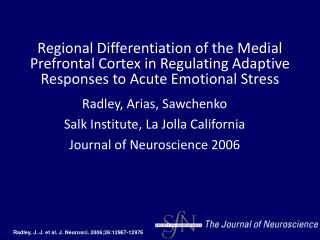
Spencer et al 2005 J. Comparative Neurology. Medial Prefrontal Ctx Control of the PVN of the Hypothalamus Response to Psychological Stress: Possible Role of the BNST……. BNST Subdivisions. Ac=anterior comm AL= anterior lateral AM= anterior medial DL= dorsal lateral Ic=internal capsule

E N D
Spencer et al 2005 J. Comparative Neurology Medial Prefrontal Ctx Control of the PVN of the Hypothalamus Response to Psychological Stress: Possible Role of the BNST……..
BNST Subdivisions Ac=anterior comm AL= anterior lateral AM= anterior medial DL= dorsal lateral Ic=internal capsule JXC=juxtacapsular LV=Lateral ventricle PL=posterior lateral PS=parastrial VL=ventral lateral VM=ventral medial
Abbreviations IIIV third ventricle ac anterior commissure ACTH adrenocorticotropic hormone ANOVA analysis of variance apPVN anterior parvocellularhypothalamic paraventricular nucleus BNST bed nucleus of the striaterminalis CeA central amygdala CRF corticotropin-releasing factor CRF-IR corticotropin-releasing factor-immunoreactivity CTb cholera toxin b subunit CTb-IR cholera toxin b subunit-immunoreactivity dBNST dorsal bed nucleus of the striaterminalis DMH dorsomedial hypothalamus dpPVN dorsal parvocellular hypothalamic paraventricular nucleus f fornix Fos-IR Fos-immunoreactivity HPA hypothalamic-pituitary-adrenal ic internal capsule IL infralimbic lpPVN lateral parvocellular hypothalamic paraventricular nucleus LS lateral septum LV lateral ventricle MeA medial amygdala mgPVNmagnocellular hypothalamic paraventricular nucleus mPFC medial prefrontal cortex mpPVN medial parvocellular hypothalamic paraventricular nucleus NTS nucleus tractussolitarius PrLprelimbic PVN paraventricular nucleus of the hypothalamus PVT paraventricular nucleus of the thalamus TH tyrosine hydroxylase vBNST ventral bed nucleus of the striaterminalis VLM ventrolateral medulla VMH ventromedial hypothalamus
Intro PVN: role in generating adaptive autonomic, behav, & hormonal stress responses Suppressive inputs are a major target for stress related issues mPFC: activation generates cardiac depressor responses, inhib influence on sympathetic vasomotor function, role in modulation of PVN….
Intro Anatomical studies fail to show direct projections from mPFC to PVN Modulation must involve one or more relay through other regions Candidate regions: medial region of hypothalamus BNST, Amygdala, paraventricular nucleus of the thalamus (PVT) LS, and brainstem catecholamine groups All of the candidate regions receive mPFC inputs and have been implicated in control of the PVN function during stress Question? Do mPFC lesions that increase PVN responses to a psychological stressor also elicit corresponding changes in activation of any of the candidates? c-fos activity
Intro • Answer! • mPFC lesion and air puff challenge causes • increased c-fos activity in vBNST, no other candidate regions • Further characterize role of BNST in PVN cell response to air puff • Think in terms of integration of info from mPFC to PVN in response to stress • Retrograde tracing to determine if BNST directly innervates PVN • Direct projections from mPFC to BNST recruited by air puff stress • Examine effects of BNST lesions on recruitment of the PVN cells to air puff stress • Determine specific regions of PVN involved
Methods • Adult male Wistar Rats • 250-500 g YIKES!!! • Individually housed, 24C 12-12 light cycle, lights on at 6 am • Food and water ad libitum • Food intake and body weight recorded throughout Surgery: Ibotenic acid used for bilateral lesions “Excitotoxicity” Death by glutamate mPFC lesions covered both prelimbic (PrL) and infralimbic (IL) regions BNST lesions covered both dorsal and ventral BNST Vascular cannulae also implanted to allow for easy collection of blood left femoral artery cannula, routed through skin up to scapulae flushed with gentamicin in heparinized saline during recovery
Methods Retrograde Tracing using the cholera toxin b subunit (CTb) ask me how this works? can be visualized using immunocytochemistry (Ctb-IR) CTb injected iontophoretically (using electric magic) into: PVN, dBNST, both d and vBNST 6 uA current for 7 seconds on and 7 seconds off, 20 minutes total Plasma ACTH conc. Determined using one giant Radioimmunoassay
Headshot from an air cannon • Series of 27 puffs of 300 kPa pressurized air aimed at the head of a freely moving animal 5-10cm away • 15 minute period • Nine blocks, each block with 3 2-second puffs with 10 seconds between puffs and 1 minute between blocks Methods Stressor!
2 separate lesions • mPFC lesion first • BNST lesion second Remember!
Substantial cell loss through all PrL and IL • No c-FOS-IR in mPFC of air puff + lesion animals Results: Characterization of mPFc lesions
C-Fos iR in coronal section of mpfc Sham Lesion mPFC Lesion
Effects of air puff on shamlesioned animals • Air puff = sig in Fos-IR in all regions • Post hoc tests on those that sig. interact w/lesion • mPFC both PrL and IL • mpPVN CRF cells • vBNST • Compared to non-stressed sham lesion control • Similar Fos profile as seen with other psychological stressors (noise, restraint) Results: Effects of sham lesions
Lesion had no effect in non-stressed animals • Table 1 sham no stress vs mPFC no stress • Lesion + stress = fos in mpPVN • Increase in the number of activated CRF cells • Sig increase in plasma ACTH with stress • No difference between sham or mPFC lesion • Lesion + stress = fos in vBNST Effects of mpfc lesion on air puff response
Sham+stress = less fos Right mpPVN mPFC lesion + stress = more fos
Fos in vBNST following mPFC lesion Sham= less fos lesion= more fos
http://ngm.nationalgeographic.com/your-shot/top-shots-2008 http://www.sciencemag.org/cgi/content/full/319/5859/101/DC1
BNST lesions vary in how much of the structure they cover BNST lesion + stress = fos compared to sham lesion + stress group Some BNST lesion + stress groups still showed high levels of fos and they were excluded from analysis as an incomplete lesion group BNST lesions
BNST sham injection BNST lesion
No effect of BNST lesion on food intake No effect on body mass BNST lesion and Food Intake
No effect in any region in non stressed groups • Table 2 sham no stress vs lesion no stress • Lesion+stress = fos in vBNST,PVN, mPFC, MeA • mpPVN showed fos • Air puff induces increased ACTH release • No sig. attenuation of the ACTH response in the BNST lesion group, despite the decreased fos expression in the CRF cells of the mpPVN Effect of BNST lesion on Stress Response
N=18: intra-PVN CTb 7d before stress • 10 have sig. tracer deposits in mpPVN • Of these 10, all showed large numbers of retrogradely labeled neurons in the vBNST • ~14% of these were co-labeled with fos • mpPVN-projecting vBNST cells sensitive to stress • No retrograde labeling in the mPFC Effect of stress on neurons projecting to PVN
Retrograde labels from CTb injections into the Mp-PVN
mPfC labels from BNST injections dBNST inj. dBNST + vBNST inj.
mPFC labeling of fos and Ctb and co-localization From BNST injections Labeling only occurred In the v and dBNST group
mPFC lesion enhanced recruitment of mpPVN CRF cells by air puff stress BNST lesion + stress = decreased fos in all regions studied mPFC and BNST important in influencing the response to a psychological stress Discussion
2 categories of acute, unconditioned stressor • Psychological: threat of a disturbance • Physical: actual disturbance of tissue integrity • Air cannon = moderate intensity • Avoids saturation of stress response • Psychological in nature (they think) • <10% total CRF cell activation in mpPVN • ~25% of what they see w/ restraint or noise stress Discussion Choice of Stressor
Air puff (along w/restraint&swim stress) elicited MeA fos activation Hemorrhage & immune challenge activate CeA Air puff in BNST activated only ventral portion Immune Challenge activates v and dBNST Air puff meets criteria of psychological stressor Discussion Choice of Stressor
mPFC lesion increases expression of fos protein by mpPVN CRF cells in response to air cannon • Not accompanied by an increase in ACTH • Could be due to altered release of other secretagogues like oxytocin or AVP • CRF release could be doing other things • mpPVN CRF also stim. NE synthesis & release in bstem Role of mpfc in CRf cell response
vBNST = increased fos following airpuff and lesion to mPFC (only candidate region that did) • vBNST gets a direct projection from mPFC, and projects directly to mpPVN • Difficult to lesion only vBNST • vBNST sends Glu to mpPVN which could be a direct stimulation following a stressor Role of BNST in mpPVN CRF cell response to air cannon
Direct projection from mPFC to ipsalateral vBNST (but not dBNST) • However, vBNST tracer injections did not retrogradely label any mPFC neurons…… • May be another brain region involved mPFC inputs to the BNST
Psychological stressor= increased mpPVN CRF and vBNST cell recruitment in mPFC lesioned animals BNST lesion = decreased mpPVN cell response to psychological stressor mPFC may suppress mpPVN CRF cell response to stress via the a BNST pathway Conclusion


![DACODA [Crandall et al.; CCS 2005]](https://cdn2.slideserve.com/4483408/dacoda-crandall-et-al-ccs-2005-dt.jpg)


![DACODA [Crandall et al.; CCS 2005]](https://cdn5.slideserve.com/9294546/dacoda-crandall-et-al-ccs-2005-dt.jpg)