Download
1 / 52
560 likes | 657 Views
X-Ray Photoelectron Spectroscopy: Theory and Practice PHYS-481 (Fall 2009). Contact Information for EMS in RRC-East. Ke-Bin Low, PhD Senior Research Specialist - Electron Microscopy Research Resources Center-East 845 West Taylor Street SES Building, Room 112 Email: kebinlow@uic.edu

E N D
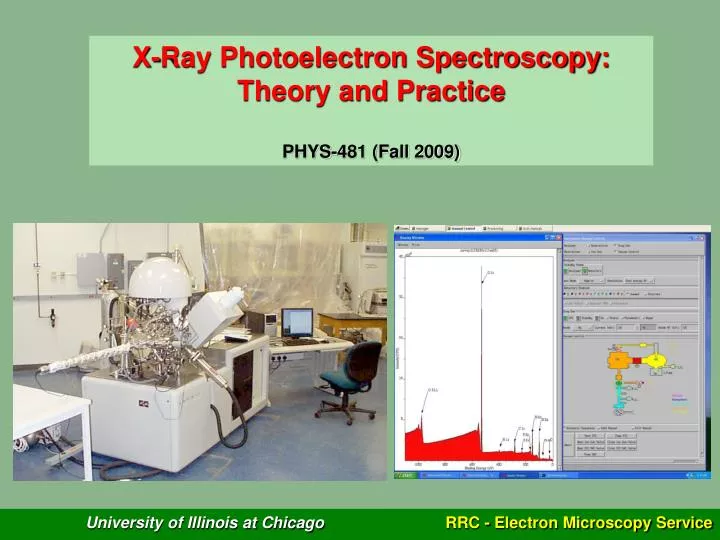
X-Ray Photoelectron Spectroscopy: Theory and Practice PHYS-481 (Fall 2009)
Contact Information for EMS in RRC-East Ke-Bin Low, PhD Senior Research Specialist - Electron Microscopy Research Resources Center-East 845 West Taylor Street SES Building, Room 112 Email: kebinlow@uic.edu Office: (312) 355-2087 Alan Nicholls, PhD Director of Research Service Facility - Electron Microscopy Research Resources Center-East 845 West Taylor Street SES Building, Room 110 Email: nicholls@uic.edu Office: (312) 996-1227
Outline of Lecture • Background • Vacuum 101 • Analytical Capabilities • Instrumentation • Spectrum Simulation • Summary
Background 1921 Photoelectric effect discovered by Albert Einstein Nobel Prize 1981 Photoemission as an analytical tool demonstrated by Kai Siegbahn (Electron Spectroscopy for Chemical Analysis – ESCA) Nobel Prize
specimen Background
Background j = |l ± s| j: Total angular momentum l: Orbital angular momentum s: Spin angular momentum
Background XPS probes core-levels → Binding energies in the range of 10 – 103 eV → Kinetic energies of similar magnitudes when Al-Kα or Mg- Kα radiation is used → Electrons with such low KE easily scattered (REMEMBER THIS)
‘Universal’ IMFP vs. KE Curve 1000 100 IMFP, λ (nm) 10 1 Kinetic Energy (eV) Background ‘Universal Curve’ shows that photoelectrons with KE in the 10 – 103 eV range have inelastic mean-free-paths (IMFPs) from 1 – 3.5 nm IMFP depends on: (1) Material (atomic #, density) (2) Kinetic energy
Background 95% of all photoelectrons detected are generated within 3λ of the surface = Sampling Depth (65% within 1λ). ‘3λ’ is used as the ‘benchmark’ definition for Sampling Depth in XPS. So the sampling depth for XPS is typically 3 - 10 nm → Surface-Sensitive! Instrument must be run under ultra-high vacuum!
Vacuum 101 VISCOUS FLOW - Whendiameter of tube > 100l, gas molecules more likely to bump into each other. Molecules in general move towards lower pressure end of tube. Unlikely to get backstreaming. MOLECULAR FLOW - When diameter of tube is < l, gas molecules are more likely to collide with the tube wall than each other. There is free movement of molecules in either direction, the numbers directly related to ratio of pressures at each end of tube. At high vacuum this ratio is likely to be close to 1. Backstreaming a concern. TRANSITIONAL FLOW - Intermediate between Viscous and Molecular.
Vacuum 101 So for an 60mm diameter tube VISCOUS > 0.1 Torr > TRANSITIONAL > 1 mTorr > MOLECULAR • OTHER PUMPS • Cryosorption - oil free, capture, Atmosphere to 10-1 Pa • Diaphragm - oil free, transfer, Atmosphere to 1Pa • Claw pump - dry, transfer, Atmosphere to 10Pa • Molecular Drag - dry, transfer, 10Pa to 10-6 Pa • Sublimation - oil free, capture, 10-1 Pa to 10-9 Pa PUMPS ION PUMP DIFFUSION ROTARY Turbo Molecular Pump • Used from 10 Pa to 10-8 Pa • Extremely high speed (10,000rpm) mechanical pumps typically with magnetic levitation bearings for EM use. • Works efficiently in Molecular Flow region - needs to be backed. • No backstreaming of oil when operating at full speed. • Major concern - preventing physical damage to pump! • Used from 1Pa to 10-7 Pa • Historically most widely used high vacuum pump, really a vapor jet pump. • Pumping speed virtually constant below 10-1 Pa • Major problem - Backstreaming; minimised by using a low vapour pressure oil. • BAD NEWS - do not let air into a diffusion pump! • Used from 10-1 Pa to 10-9 Pa • Gas molecules that are pumped are trapped inside pump by the gettering action of the sputtered Ti - limited lifetime. • Absolute freedom from oil contamination with no moving parts. • Ideal for high vacuum systems but are not well suited on systems that are cycled frequently to atmosphere. • Used from atmosphere down to 0.1Pa • Problems with corrosive of condensable gases (H2O) • Potential source of oil contamination of vacuum system if pressure in line to system is not kept in viscous flow regime. • At or near atmospheric pressure an oil mist is ejected through outlet valve. Must vent outside or through a filter. REMEMBER -- No Pump exerts a force that drags or pulls gas molecules to it. Pumping is purely diffusion of gas molecules from high pressure to low pressure regions
Analytical Capabilities of XPS • Identify elements/compounds (except H and He) • Determine oxidation states (e.g. Ti3+ or Ti4+) • Identify types of chemical bonds (e.g. Si-O or Si-C) • Semi-quantitative analysis (10-15% error) • Determine adsorbate/film thickness • Highly surface-sensitive (3 – 10 nm from the surface) • → Detection limit 0.1 to 1 at% • → Ultra-high vacuum required!!! • → Minimize/delay surface reactions and contaminations
KINETIC ENERGY, eV Analytical Capabilities of XPS Survey spectrum for element identification
Analytical Capabilities of XPS XPS spectra show characteristic "stepped" background. Due to inelastic processes (extrinsic losses) from deep in bulk. Electrons deeper in surface loose energy and emerge with reduced KE, apparent background increase at higher BE
Analytical Capabilities of XPS • Typical Features and ‘Artifacts’ of Core-Level Peaks • Can get multiple peaks from core levels – must be aware of where they • come from in order to carry out chemical analysis (not all may be present) • Spin orbit splitting leads to additional peaks (no splitting for s, splitting for p,d,f etc.) • Additional peaks due to, for example, chemical shifts and oxidation states • Ghost peaks at lower binding energies (achromatic X-ray only)– no useful info! • Shake up/ off peaks at higher binding energies (result of energy being transferred from the ejected photoelectron electron to a valence electron). • Plasmon loss peaks (due to electron excitations) • Photon-induced Auger peaks • Effects of charging of non conductive specimens
O 1s 537.0 534.0 531.0 528.0 525.0 Binding Energy (eV) Analytical Capabilities of XPS No spin-orbital splitting for s Spin-orbital splitting for p, d, f
Analytical Capabilities of XPS Ti 2p1/2 and 2p3/2 chemical shift for Ti and Ti4+. Charge withdrawn Ti → Ti4+ so 2p orbital relaxes to higher BE
Analytical Capabilities of XPS Schematic of Ghost and Shake-up peaks Ghost Peak Shake-up Peak Main peak Binding Energy Kinetic Energy
Analytical Capabilities of XPS Electrical insulators cannot dissipate charge generated by photoemission Process. Surface picks up excess positive charge - all peaks shift to higher BE Can be reduced by exposing surface to neutralizing flux of low energy electrons - "flood gun" or "neutralizer“. BUT must have good reference peak.
Analytical Capabilities of XPS Is = Io exp (-d / λcosθ) Is: Intensity at surface Io: Intensity from infinitely-thick sample d: depth λ: Inelastic mean-free-path (IMFP) θ: Spectrometer take-off angle Beer-Lambert relationship (Numerical expression Describing the photoelectron Intensity generated from a material)
SiO2 Surface Layer Si Substrate Analytical Capabilities of XPS Using the Beer-Lambert expression to estimate film thickness…
When λ, θ and all the respective intensities are known, film thickness can be determined by taking the ratio of Ifilm to Isubstrate and solve for d ISi: 1411.6 ISiO2: 241.4 Analytical Capabilities of XPS
Analytical Capabilities of XPS Semi-Quantitative Analysis Photoelectron intensity from a homogeneous material is also dependent on instrumental factors, and can be alternatively-described by I = JCσζTλ J: X-ray flux C: Concentration of the element-of-interest σ: Ionization cross-section ζ: Spectrometer angular acceptance T: Spectrometer transmission function λ: IMFP of the element
Analytical Capabilities of XPS • C = I/(JσζTλ) = I/JF • F is termed the atomic sensitivity factor: • - incorporates all the terms associated with the spectrometer and material • empirically-determined by XPS manufacturer • values are normalized against Fluorine
Analytical Capabilities of XPS • Atomic fraction of an element (A) in a multi-component material (ABCD…) can be estimated using the following, • Atomic % A = (IA/FA) / S (In/Fn) • Quantification using this expression is valid only if: • Material is homogeneous, • Material surface is smooth and flat.
Charge Neutralizer (built into lenses) Ion Gun Instrumentation
Instrumentation Kratos Axis-165 XPS system in RRC-East
100 % Total FWHM Intensity 50 % Energy Instrumentation Total resolution (i.e. peak’s full-width-half-maximum) of instrument is convolution of: (1) X-ray energy spread, (2) Spectrometer broadening, and (3) Intrinsic line-width of the element-of-interest. Total FWHM = {FWHMx-ray2 + FWHMspectrometer2 + FWHMintrinsic2}1/2
Instrumentation Binding energy shifts due to different chemical states or bonding configurations can be subtle (1eV or less) for certain elements. Say, if achievable total FWHM of a peak is 2 eV, the XPS instrument will not be able to resolve 2 peaks that are separated only by 0.5eV!!! Total FWHM can only be decreased by minimizing the FWHM of: (1) X-ray; (2) Spectrometer * Intrinsic line-widths is a non-controllable term!
Instrumentation • Ideal Candidates for X-ray source: • Al- and Mg-Kα • High energy • (2) Narrow energy spread
Instrumentation Twin-Anode Achromatic X-ray source: Bombard metallic anode with 10-25kV electrons with ~10mA of current to generate X-rays. Can generate high X-ray flux producing high signal BUT specimen may be damaged by heat generated by the X-rays and continuum radiation and source emits X-ray satellites (additional weak lines at lower binding energies) Simple, relatively inexpensive High flux (1010-1012 photons·s-1) Beam size ~ 1cm
Electron Spectrometer Quartz Crystal Sample RowlandCircle X-ray Source Instrumentation Monochromatic X-Ray source: Diffraction from bent SiO2 crystal focusing primary λ at specimen. Other λ's focused at different points in space (filtered). Always use Al Kα which is diffracted from quartz (no equivalent crystal for Mg Kα) Beam size ~ 1 cm to 50 mm Eliminates satellites peaks – simpler spectra Decreases FWHM of X-ray energy Flux decreases at least an order of magnitude leading to less damage, improves S/N (no X-ray continuum) but lower signal More complicated and expensive
Instrumentation Most common type of electrostatic deflection-type analyzer: Concentric Hemispherical Analyzer (CHA) or spherical sector analyzer Energy resolution dependant on radius. Capable of collecting photoelectrons of larger angular distribution. Photoelectrons of a specific energy are focused by the lens at the slit of the spectrometer. Lens also controls sampling area. Photoelectrons travel through a circular path and exit into a series of channeltrons (electron multipliers).
Instrumentation An electron of kinetic energy eV = Vo will travel a circular orbit through hemispheres at radius Ro Since Ro, R1 and R2 are fixed, in principle changing V1 and V2 will allow electrons of different KE to be detected. Negative potential on two hemispheres V2 > V1 Potential of mean path, Ro through analyzer is Vo = (V1R1+V2R2)/2Ro
Peak FWHM Photoelectron KE Spectrometer Term (constant) Instrumentation But DE/Eo = S/2Ro DE = (S/2Ro)Eo Spectrometer broadening (i.e. FWHMspectrometer) is a function of photoelectron KE entering spectrometer… i.e. Resolution is non-uniform across XPS energy spectrum!
Instrumentation Single-Channel Electron Multipliers (Channeltron) - Hollow glass tube with semiconducting layer on inner surface - Electrons/ions entering Channeltron produce secondary electrons (SEs) - ‘Avalanche’ effect as SEs accelerate down the tube under HV - Signal amplified by ~ 106 to 108
Instrumentation To circumvent the problem… (1) Hemisphere potentials are fixed to allow electrons with a fixed KE (the pass energy,Ep) to reach the electron detectors (Channeltrons); (2) Electrostatic lens before the slit decelerate photoelectrons of a particular KE to Ep; (3) Magnetic lens focus these electrons with Ep at the slit, so that only electrons with that pass energy are allowed to enter the slit. Scanning of the energy-scale is achieved by varying the decelerating potential on the electrostatic lens instead of the hemisphere potentials → Fixed energy-resolution across the energy-scale!!! Something to Ponder… Should an infinitesimally-small pass energy yield an extremely mild spectrometer broadening???
Instrumentation • Most XPS systems also come equipped with an ion gun. • Purpose: • Removes surface contaminants (usually oxides and hydrocarbons); • Allows depth-profiling study. • How does it ‘clean’ a surface? • Ionizes an inert gas (usually Ar) • Focuses and accelerates the ions towards the specimen surface • Rasters the ion beam across the surface • Ions impart energy to surface contaminants → Sputtering them away!
Instrumentation Example: Successful removal of Cr-contamination from CdTe
Instrumentation • Potential Problems Associated with Sputter-Cleaning: • Surface roughening; • Alter oxidation states of some elements; • Selective ‘etching’ of surface due to different sputtering rates in a multi-component material.
Instrumentation Example: Oxidation-state changed in Pd after sputtering…
Spectrum Simulation (Peak-Fitting) • Simulation of XPS spectra (i.e. peak fitting) to match experimentally-observed spectra. • Purpose: • Background noise subtraction to reveal true peak intensities; • Precise determination of peak position; • Deconvolute spectra into individual components when 2 or more peaks are in close proximity.
Spectrum Simulation (Peak-Fitting) Select Background Model Background Quality? Modify Peak Parameters Poor Good Option 1 Adding Synthetic Peak Option 2 Poor • Providing Initial-Guess • For Peak Parameters: • Line position • Area • FWHM • Gaussian-Lorentzian Ratio • Asymetry • S.O.S. End of Fitting Software Fitting Fit Quality? Good
Spectrum Simulation (Peak-Fitting) Linear • Types of Background: • Linear • Shirley (typically-used) • Tougaard Tougaard Shirley
Spectrum Simulation (Peak-Fitting) • Types of Line-Shapes: • Gaussian Function • → Describes the measurement process • (e.g. instrumental response, X-ray line-shape, Doppler and thermal broadening) • Lorentzian Function • → Describes lifetime broadening • (intrinsic line-width) • XPS peaks can usually be described by varying G-L ratio
Spectrum Simulation (Peak-Fitting) Peaks associated with pure metals may tend to be asymmetic… → Need to introduce a ‘Tail Modifier’ term to the G-L functions
Spectrum Simulation (Peak-Fitting) XPSPEAK version 4.1 is a free windows-based XPS peak fitting program available online. Installation file and user manual downloadable at: http://www.phy.cuhk.edu.hk/~surface/XPSPEAK You will need this software to complete the XPS assignments!