Download
1 / 16
160 likes | 198 Views
This lecture explores the neurophysiological basis of movement in the spinal cord, focusing on the laminar structure, excitatory and inhibitory synapses, and the role of recurrent and reciprocal inhibition.

E N D
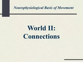
Neurophysiological Basis of Movement World II: Connections
Back Front I II III IV V VI VII X VIII IX Lecture 7: Excitation and Inhibition Within the Spinal Cord The spinal cord has a laminar structure. It “flows” along the body, preserving the general picture of its cross-sections (upper figure). At each level, the gray matter forms a characteristic butterfly picture consisting of 10 Rexed’s laminae (lower figure).
Rostral Dorsal Medial Dorsal Ventral Lateral Proximal Caudal Ventral Distal Geographical Terms These geographical terms will be helpful in future descriptions. The figure of a person is drawn in a sagittal plane.
Spinal cord Spinous process Vertebra Dorsal root Ventral root Spinal Roots Each vertebra has a body and a spinous process. Peripheral information gets into the spinal cord through the dorsal roots, while efferent signals are sent from the spinal cord through the ventral roots.
C1 C1 Spinal cord Vertebrae C8 C7 T1 T1 Ventral roots Dorsal roots T12 L1 T12 L5 S1 L1 S5 L5 Cauda Sacrum equina Vertebrae and Segments Starting from the skull, vertebrae are numbered from C1 to L5 and then end with the sacrum. Spinal segments are numbered from C1 to S5, but this classification does not correspond exactly to the vertebrae classification. The spinal cord ends at the L1 vertebra; lower, the roots of the lower segments form the cauda equina.
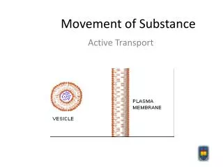
Synaptic vesicles Presynaptic membrane Synaptic cleft Postsynaptic membrane Excitatory and Inhibitory Synapses A synapse consists of a presynaptic membrane, a synaptic cleft, and a postsynaptic membrane. Inhibition means a decrease in the efficacy of the synapse and may occur as a result of events on the presynaptic or the postsynaptic membrane.
Excitatory synapse Inhibitory synapse V V Time Time Threshold EPSP IPSP EPSP and IPSP An excitatory synapse leads to a depolarization of the postsynaptic membrane (i.e., bringing it closer to the threshold), whereas an inhibitory synapse leads to a hyperpolarization of the postsynaptic membrane.
-MN a -MN a -MN g a Renshaw cell Muscle Recurrent Inhibition Axons of a-motoneurons branch very close to the cell body and make excitatory synapses on Renshaw cells. These cells, in turn, make inhibitory synapses on a-motoneurons of the same pool as well as on -motoneurons, sending their axons to spindles in the same muscle.
Reciprocal Inhibition Agonist pool Ia IN Ia interneurons receive excitatory inputs from Ia afferents and make inhibitory synapses on motoneurons innervating the antagonist muscle. Ia INs are inhibited by Renshaw cells and also receive descending inputs. Ganglion -MN a Antagonist pool -MN a Renshaw cell T-shaped axon Ia afferent Antagonist muscle Agonist muscle
_ + Ia INs -MNs a Ia afferents + + + Agonist Agonist force force Antagonist length _ _ Renshaw -MNs a Ia INs -MNs a cell + + + Antagonist force Schemes of Recurrent and Reciprocal Loops Feedback loops involving Ia interneurons and Renshaw cells. A plus sign means that an increase in the input leads to an increase in the output. A minus sign means that an increase in the input leads to a decrease in the output.
Recurrent and Reciprocal Inhibition Recurrent inhibition: • Alpha-motoneurons of a pool fire. • They send axon branches to small inhibitory interneurons (Renshaw cells) in the ventral horns of the spinal cord. • Renshaw cells inhibit all motoneurons of the same pool. Reciprocal inhibition: • Small interneurons (Ia interneurons) are activated by primary spindle afferent fingers (Ia afferents). • The Ia interneurons inhibit motoneurons of the antagonist muscle.
Inhibitory synapse Postsynaptic Inhibition A postsynaptic inhibitory synapse hyperpolarizes the postsynaptic membrane and decreases its responsiveness to excitatory synapses (shown by open circles).
Presynaptic membrane Synaptic cleft Postsynaptic membrane Presynaptic inhibition Presynaptic Inhibition Presynaptic inhibition acts selectively on certain synapses. It involves an excitatory synapse acting on the presynaptic membrane, inducing its steady subthreshold depolarization, thus decreasing the amount of mediator released in response to a single presynaptic action potential.
Neurotransmitter A NT AAP A Voltage B Time AAP Threshold VEQ What Makes Presynaptic Inhibition Possible? A: A relatively small change in the peak-to-peak amplitude of the presynaptic action potential (A) leads to a major change in the amount of released neurotransmitter (NT). B: A small, steady depolarization leads to a drop in the peak-to-peak AP amplitude and a decrease in the synapse efficacy.
Presynaptic inhibition a-MN Afferent fiber Muscle An Example of Presynaptic Inhibition In this figure, an excitatory synapse is acting at a presynaptic afferent (sensory) fiber close to its synapse at the target -motoneuron.
Current PIC PIC Voltage −70 PIC The Role of Persistent Inward Currents (PICs) An illustration of weak effects (solid line), moderate effects (dashed line), and strong effects (dotted line) of persistent inward currents (PIC).