Download

1 / 33
360 likes | 1.03k Views
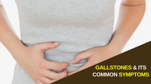
GALLSTONES AND ITS COMPLICATIONS. PATHOGENESIS. RISK FACTORS. A ge: Increasing age* B ody habitus: Obesity, rapid weight loss C hildbearing: Pregnancy

E N D
RISK FACTORS • Age: Increasing age* • Body habitus: Obesity, rapid weight loss • Childbearing: Pregnancy • Drugs: Fibric acid derivatives (or fibrates), contraceptive steroids, postmenopausal estrogens, progesterone, octreotide (Sandostatin), ceftriaxone (Rocephin) • Ethnicity: Pima Indians, Scandinavians • Family: Maternal family history of gallstones • Gender: Females • Hyperalimentation: Total parenteral nutrition,† fasting • Ileal and other metabolic diseases: Ileal disease (Crohn's disease), resection or bypass,* high triglycerides, diabetes mellitus, chronic hemolysis,* alcoholic cirrhosis,* biliary infection,* primary biliary cirrhosis, duodenal diverticula,* truncal vagotomy, hyperparathyroidism, low level of high-density lipoprotein cholesterol • *--Risk factors for pigment gallstone formation.†--Risk factor for cholesterol and pigment gallstone formation.
Case • 46yo F w RUQ pain x4hr, after a fatty meal, radiating to the R scapula, also w nausea. Pt is pain-free now. • No prior episodes • Minimal RUQ tenderness, no Murphy’s • WBC 8, LFT normal • RUQ U/S reveals cholelithiasis without GB wall thickening or pericholecystic fluid
CASE • → denotes gallstones • ► denotes the acoustic shadow due to absence of reflected sound waves behind the gallstone → → ►
Symptomatic cholelithiasis • aka “biliary colic” • The pain occurs due to a stone obstructing the cystic duct, causing wall tension; pain resolves when stone passes • Pain usually lasts 1-5 hrs, rarely > 24hrs • Ultrasound reveals evidence at the crime scene of the likely etiology: gallstones • Exam, WBC, and LFT normal in this case • Treatment: Laparoscopic cholecystectomy
CASE • 66 years-old male with past medical history of diabetes mellitus, c/o RUQ pain > 24hrs radiating to the R scapula, started after fatty meal, a/w nausea, vomiting, fever • Exam: Palpable, tender gallbladder, guarding, +Murphy’s = inspiratory arrest • WBC 18, Mild ↑LFT
Case • Curved arrow • Two small stones at GB neck • Straight arrow • Thickened GB wall • ◄ • pericholecystic fluid = dark lining outside the wall ◄
U/S: gallstones, wall thickening (>4mm), GB distension, pericholecystic fluid, sonographic Murphy’s sign (very specific) • Diagnosis: ?
CASE • A 72 year old woman with heart faliure, hypertension and diabetes came to the office with a chief complaint of chills, abdominal pain, nausea and vomiting followed by inability to pass flatus 8 hours prior to consult. • On physical examination the patient was febrile and appeared dehydrated. Abdominal exam: distension and increased bowel sounds. No jaundice. • Labs: Hyperglycemia, mild renal failure, hypernatremia and leukocytosis.
A 43-year-old woman visited our outpatient clinic in March 2004 with a six-month history of colic pain, nausea and vomiting. She had a past history of hyperthyroidism. Physical examination revealed a tender right upper abdomen and jaundice. The laboratory blood tests showed increased levels of bilirubin 9 mg/dl alkaline phosphatase 145 U/l (<140 U/l), g-glutamyltransferase 161 U/l (<40 U/l), alanine transaminase 1240 U/l (<40 U/l), aspartate transaminase 688 U/l (<35 U/l) and lactate dehydrogenase 888 U/l (<480 U/l), while the serum amylase was normal. Ultrasonography showed slightly dilated intrahepatic ducts, an extended gallbladder with multiple stones CASE
One study has shown that acute cholecystitis resolves without complications in approximately 83% of patients but results in gangrenous cholecystitis in 7%, gallbladder empyema in 6%, perforation in 3%, and emphysematous cholecystitis in less than 1% • When the level of leukocytosis exceeds 15,000 cells/mm, particularly in the setting of worsening pain, high fever (temperature >102 °F), and chills, suppurative cholecystitis (empyema) or perforation should be suspected, and urgent surgical intervention may be required.
CASE • 46yo F p/w RUQ pain, jaundice, acholic stools, dark tea-colored urine, no fevers • Known history of cholelithiasis • Exam: unremarkable • WBC 8, T.Bili 8, AST/ALT 90/75, HepB/C neg • Ultrasound: Gallstones, CBD not dilated • Diagnosis: ?
MRCP EUS CASE
CHOLEDOCHOLITHIASIS • Fifteen percent of patients with gallbladder stones also have CBD stones. Conversely, of patients with ductal stones, 95% also have gallbladder stones • Stones in the CBD usually come to rest at the lower end of the ampulla of Vater. Obstruction of the bile duct raises bile pressure proximally and causes the ducts to dilate. Pressure in the CBD is normally 10 to 15 cm H2O and rises to 25 to 40 cm H2O with complete obstruction. When pressure exceeds 15 cm H2O, bile flow decreases, and at 30 cm H2O, bile flow • The bile duct dilates to the point that it can be detected on either ultrasonography or abdominal CT in approximately 75% of cases. In the patient who has had recurrent bouts of cholangitis, the bile duct may become fibrotic and thus unable to dilate. Moreover, dilatation of the duct is sometimes absent in patients with choledocholithiasis because the obstruction is low-grade and intermittent
CHOLEDOCHOLITHIASIS • Acute obstruction usually causes biliary pain and jaundice Obstruction that develops gradually over several months may manifest initially as pruritus or jaundice alone. If bacteria proliferate, life-threatening cholangitis may result • The physical findings are usually normal if obstruction of the CBD is intermittent. Mild to moderate jaundice may be noted when obstruction has been present for several days to a few weeks. Deep jaundice without pain, particularly with a palpable gallbladder (Courvoisier's sign), suggests neoplastic obstruction of the CBD, even when the patient has stones in the gallbladder. With long-standing obstruction, secondary biliary cirrhosis may result, leading to physical findings of chronic liver disease.
CHOLEDOCHOLITHIASIS • Elevated serum bilirubin and alkaline phosphatase levels are seen with CBD obstruction Serum bilirubin level >10 mg/dL suggests malignant obstruction or coexisting hemolysis A transient “spike” in serum aminotransferase or amylase (or lipase) levels suggests the passage of a stone
CASE • 46yo F p/w fever, RUQ pain, jaundice • If also altered mental status and signs of shock • VS tachycardic, hypotensive • ABC’s, Resuscitate • 2 large bore IV, Foley, Continuous monitor • 1-2L fluid bolus, repeat until resuscitated • Diagnosis: ?
CHOLANGITIS • Stone in the common bile duct causing bile stasis Bacterial superinfection of stagnant bile Early bacteremia • Charcot's triad (pain, jaundice, and fever) is present in 70% of patients Pain may be mild and transient and is often accompanied by chills Mental confusion, lethargy, and delirium suggest sepsis • Fever in 95% RUQ tenderness in 90% Jaundice in 80% Peritoneal signs in 15% Hypotension and mental confusion coexist in 15% and suggest gram-negative sepsis • Leukocytosis in 80%, but remainder may have normal white blood cell count with band forms Serum bilirubin level >2 mg/dL in 80% Serum alkaline phosphatase level is usually elevated Blood cultures are usually positive, especially during chills or fever spike; two organisms are grown in cultures from one half of patients
CASE • 46yo F w RUQ pain x4hr, after a fatty meal, radiating to the R scapula, also w nausea. Pt is pain-free now. Has had multiple prior attacks of similar RUQ pain • No prior episodes • Minimal RUQ tenderness, no Murphy’s • WBC 8, LFT normal • RUQ U/S reveals cholelithiasis with GB wall thickening, no pericholecystic fluid
CHRONIC CHOLECYSTITIS • Chronic inflammation of the gallbladder is the indication for nearly 3% of cholecystectomies in adults. • Pathophysiology is poorly understood. repeated episodes of low-grade gallbladder obstruction, resulting in recurrent mucosal trauma . • There is little correlation between the number of choleliths or their overall volume and the degree of gallbladder wall inflammation. • In fact, 12% to 13% of patients who have chroniccholecystitis have no demonstrable stones. • No role of Bacterial infection of the bile • As each episode of acute inflammation resolves, neutrophilic infiltration is replaced with lymphocytes, plasma cells, macrophages, and eosinophils. Focal ulcerations and necrotic tissue are replaced by granulation tissue and collagen deposits. The gallbladder wall may become thickened or remain thin. The mucosa can remain intact, develop accentuated folds, or be flattened.
The symptoms of chronic cholecystitis • vary : classic severe biliary colic vague or nonspecific complaints. intermittent episodes of nausea, reflux symptoms, food intolerance, or bloating. low-grade fever, mild upper abdominal discomfort, or chronic fatigue. • Not infrequently patients who have chroniccholecystitis have been treated for gastritis, ulcer disease, or irritable bowel syndrome without appreciable improvement in their complaints. • HIDA with CCK helps
Take Home Points • As always, ABC & Resuscitate before Dx • Understanding the definitions is key • Is this acute cholecystitis? (fever, WBC, tender on exam with positive Murphy’s) • Or simply cholelithiasis vs ongoing chronic cholecystitis? (no fever/WBC) • Is patient sick or toxic-appearing, to suspect empyema, gangrene or even perforation? • Elicit h/o jaundice, acholic stools, tea-colored urine • Rule out cholangitis, because this will kill the patient unless dx & tx early