Download
1 / 44
450 likes | 681 Views
Digestion : Day 2. Another Look at the Bolus. Visceral smooth muscle shows rhythmic cycles of activity Pacemaker cells ( these set up your BASIC ELECTRICAL RHYTHM (BER) . These cells are called the “interstitial cells of cajal”. Set up a constant rate of peristaltic wave of 3 per minute.

E N D
Another Look at the Bolus • Visceral smooth muscle shows rhythmic cycles of activity • Pacemaker cells( these set up your BASIC ELECTRICAL RHYTHM (BER) . These cells are called the “interstitial cells of cajal”. Set up a constant rate of peristaltic wave of 3 per minute. • Peristalsis • Waves that move a bolus • Segmentation • Churn and fragment a bolus
Pharynx • 3 Parts: • Nasopharynx: behind the nasal cavity • Oropharynx: extends from the soft palate to the epiglottis. • Laryngopharynax: extends from the epiglottis to the base of the larynx, which is continous w/ the esophagus. • Function: Initiate peristalsis
Esophagus • Extends from pharynx through the diaphragm to the stomach. • Continues peristalsis • A thickening of the smooth muscle , the esophageal sphincter, at the esophageal-stomach junction, controls the food passage into the stomach.
STOMACH • Left side of abdominal cavity • Different Regions:1. Cardiac (Opening), 2.Fundus (Expanded portion,lateral to cardiac), 3.Body( Midpart) and 4.Pylorus (Terminal part of the stomach which is continuous w/ the sm. Intest.) • Function(s): 1. Temporary Storage Area 2. Site for chemical digestion of Protein. • ALSO, the Glands are going to be different for the various sections……..
Figure 24.12 The Stomach Figure 24.12b
Cells of Gastric Glands • Mucous cells secrete mucus • Regenerative cells • divide rapidly to produce new cells that migrate to surface • Parietal cells • secrete HCl acid and intrinsic factor • Chief cells • secrete pepsinogen • chymosin and lipase in infancy • Enteroendocrine cells • secrete hormones and paracrine messengers • D and G cells
What is a ZYMOGEN? • PEPSINOGEN is one. • It is an inactive enzyme, usually a long molecule that, when shortened, becomes activated. • Pepsinogen is activated by hydrochloric acid as it cleaves Pepsinogen to PEPSIN.
DIGESTION OF NUTRIENTS • Carbohydrates: Oral Cavity • Small Intestines • Proteins: Stomach • and Small Intestines • Fats: Small Intestines
Carbohydrates Mouth: Amylase( present in saliva) digests starch to oligosaccharides. Has pH optima of 6.75 -7.0 Sm. Intestines: Pancreatic amylase; oligosaccharides to dissacharide (maltose) Brush border enzymes: (dextrinase and glucoamylase): hydrolyze dissacchrides to monosaccharides. (these enzymes are secreted from the duodenal mucosum)
Proteins • Digestion starts in Stomach with PEPSIN ( optimum pH 1.5-2.5) Polypeptides reduced to oligopeptides • Small Intestine with Pancreatic enzymes TRYPSIN and CHRYMOTRYPSIN into smaller peptides ( 2 or 3 amino acids) • Brush border enzymes (aminopeptidase and dipeptidase finalize digestion to amino acid monomers
About Lipid Digestion • Begins and is Finished in the Small Intestines. • Enzyme? Just say Lipase for Triglycerides • Triglycerides that are not digested, combine w/ cholesterol and phospholids to form chylomicrons “milky fat” ; These are too large to enter capillaries and enter the lacteal. They form milly portion of lymph. • Eventually this extra fatty lymph will empty into blood stream via thoracic duct. • LIPOPROTEIN LIPASE will digest to fatty acids and glycerol or new chylomicrons are used to transport cholesterol back to liver.
Nucleic Acid Digestion • For Both DNA and RNA. • To nucleotide monomers • In the Small Intestine only. • By pancreatic enzyme and Brush Border (nucleosidases and phosphatases)
Digestion and absorption in the stomach • Preliminary digestion of proteins (process finished in the small intestine) • Pepsin from pepsinogen • Very little absorption of nutrients . • Some drugs, however, are absorbed. • Mucous secretion containing several hormones • Enteroendocrine cells ( means hormone secreting cells) • G cells secrete gastrin • D cells secrete somatostatin
What is SOMATOSTATIN? • This is also called Growth Hormone Inhibiting Hormone (GHIH) • Released from stomach mucosa ( D cells) • Inactivates gastric secretions • Inhibits gastric motility and emptying • D cells are activated by low pH (when stomach is EMPTY)
Small intestine • Important digestive and absorptive functions • Secretions and buffers provided by pancreas, liver, gall bladder • Three subdivisions: • Duodenum • Jejunum • Ileum • Ileocecal sphincter • Transition between small and large intestine
Figure 24.16 Regions of the Small Intestine Figure 24.16a
Histology of the small intestine • Plicae Circulares • Transverse folds of the intestinal lining (Deep folds in mucosal and submucosal layers…this allows chyme to stay in SI longer ..more time form digestion. • Villi • Fingerlike projections of the mucosa • Lacteals • Terminal lymphatic in villus • Intestinal glands • Lined by enteroendocrine, goblet and stem cells
Figure 24.17 The Intestinal Wall Figure 24.17b, c
Some Terms To Review • Microscopic Anatomy: • A villus in Small Intestine is covered with two kinds of epithelial cells; Absorptive and Goblet. • Goblet: Secrets mucus • Absorptive; It is for absorption; This is your BRUSH BORDER CELL
Intestinal juices • Moisten chyme • Help buffer acids • Maintain digestive material in solution
Small Intestine • SPECIFIC TO CERTAIN SI REGIONS: • Duodenum: (Brunner’s glands) • Produce a mucus that 1. Alkaline mucus: buffers acidity protects epithelial lining of GI tract from erosion; 2.Enterokinase which activates trypsinogen to trypsin and 3. Urogastrone,:inhibits secretion of hydrochloric acid in stomach. • Ileum • aggregated lymphoid nodules (Peyer’s patches)
Intestinal movements • Peristalsis • Segmentation ( • Gastroenteric reflexes • Initiated by stretch receptors in stomach • Gastroileal reflex • Triggers relaxation of ileocecal valve
Lipolysis- Lipids are digested chemically in the small intestine to fatty acids and glycerol. The lipid we see most in the diet is the triglyceride which consist of one molecule of glycerol and 3 fatty acids. Digestion in The Small Intestine
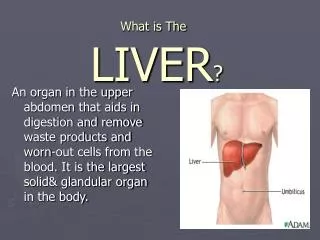
Before Fat is Chemically Digested it is Emulsified By Bile. • LIVER: (produces Bile) and secreted through bile canliculi. Leave through common hepatic duct toward duodenum. • Gallbladder:(Stores extra bile) • Bile enters duodenum when gallbladder contracts. • Stimulus from GB contraction is a hormone…CCK. Vagus nerve is also a minor control. • Hepatopancreatic sphincter opens and bile and pancreatic juice and enter SI
Figure 24.21 The Gallbladder Figure 24.21a, b
The Fat DIGESTIVE ENZYMES • LIPASES : Fat-digesting enzymes • Large fats are first broken down mechanically, to smaller fats and “pretreated” with bile salts ( which have a polar and non-polar region. Now, fats are smaller and can be suspended in hydrophilic solution (MORE SURFACE AREA) • End Products:1) Fatty Acids and Glycerol ( these go into blood stream (diffusion through villi)2) chylomicron( Actually are protein and fat together, you know these. They can be similar to the HDLs and LDLs. ANYWAY the are found during the end of LIPID digestion. They are absorbed into the lacteal. Remember, lymph is composed partially of lipids; this is the one. Chylomicron means MILKY-FAT.
Water Balance • Digestive tract receives about 9 L of water/day • .7 L in food, 1.6 L in drink, 6.7 L in secretions • 8 L is absorbed by small intestine and 0.8 L by large intestine • Water is absorbed by osmosis following the absorption of salts and organic nutrients • Diarrhea occurs when too little water is absorbed • feces pass through too quickly if irritated • feces contains high concentrations of a solute (lactose)
Gross Anatomy of Large Intestine • 5 feet long and 2.5 inches in diameter. • Begins as cecum and appendix in lower right corner • Ascending, transverse and descending colon frame the small intestine • Sigmoid colon is S-shaped portion leading down into pelvis • Rectum - straight portion ending at anal canal
Bacterial Flora and Intestinal Gas • Bacterial flora populate large intestine • ferment cellulose and other undigested carbohydrates; we absorb resulting sugars • synthesize vitamins B and K • Flatus (gas) • average person produces 500 mL per day • most is swallowed air but hydrogen sulfide, indole and skatole produce odor
Bacterial Flora and Intestinal Gas • Bacterial flora populate large intestine • ferment cellulose and other undigested carbohydrates; we absorb resulting sugars • synthesize vitamins B and K • Flatus (gas) • average person produces 500 mL per day • most is swallowed air but hydrogen sulfide, indole and skatole produce odor
Absorption and Motility • Transit time is 12 to 24 hours • reabsorbs water and electrolytes • Feces consist of water and solids (bacteria, mucus, undigested fiber, fat and sloughed epithelial cells • Haustral contractions occur every 30 minutes • distension of a haustrum stimulates it to contract • Mass movements occur 1 to 3 times a day • triggered by gastrocolic and duodenocolic reflexes • filling of the stomach and duodenum stimulates motility • moves residue for several centimeters with each contraction