Download
1 / 25
300 likes | 1.26k Views
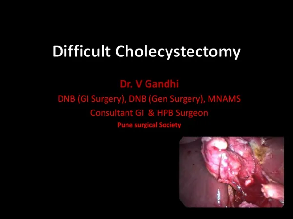
Bill Cavatassi. Complications During A Laparoscopic Cholecystectomy. Outline. Overview Bleeding Pneumoperiteneum creation Spilled stones Bile Injury. Overview. 750,000 per year 90% of all CCY are lap 25% of surgeons with pneumo creation complication

E N D
Bill Cavatassi Complications During A Laparoscopic Cholecystectomy
Outline Overview Bleeding Pneumoperiteneum creation Spilled stones Bile Injury
Overview 750,000 per year 90% of all CCY are lap 25% of surgeons with pneumo creation complication 34-49% of surgeons will have a bile duct injury
Major complications 2.6% Bleeding (0.11 – 1.97) Abscess (0.14 – 0.3) Bile leak (0.3-0.9) Bile injury (0.26 – 0.6) Bowel injury (0.14 – 0.35) Gas embolism (0.001%)
Variable Odds Ratio AC vs CC-------------- Male vs female------- Age---------------------- Body weight----------- Experience------------ Time-------------------- 1.86 1.18 1.12 per 10 years 1.25 (>90 vs 60-89) 1.34 (>90 vs <60) 1.22 (>100 vs <10) 1.36 (>100 vs 11-99) 90% in first 30 cases 1.68 for each 30 mins >2hrs 4x one hr case
Bleeding (0.07-1.9%) Sites: liver, arterial sources, port insertion sites) Liver: removal GB from fossa Lap hemostasis –vs- open stitch ligature (8%) Arterial source during resection usually cystic artery – clip if anatomial landmarks ensured
Pneumo Creation Carbon dioxide – colorless, noncombustible, inexpensive Rapid absorption (good: dec gas embolism, bad: hypercarbia) Venous Return: decrease Esp if hypovolemic, due to cava compression Heart Rate: increase Hypercarbia causes tachycardia and PVCs Peritoneal inflammation can cause vagal response and dec HR CVP: usually artificially elevated from inc thoracic pressures
FRC (functional reserve capacity): decreases Diaphragm motion limited, inc peak airway pressures to maintain same tidal volume MAP and SVR: increase CO: decrease (inc MAP and dec venous return) GFR and urine output: decrease Dec renal vein blood flow pH: decrease Hypercarbia leading to respiratory acidosis No adverse effects in healthy pts, corrected with inc minute ventilation Problem in COPD because dec ability to get rid of CO2 (intermittent ABGs)
Complications from pneumoperitoneum creation Mortality 0.2% Incidence of injury 0.2% Veress & Hasson Only 60% dx’d at time of injury
Retro of 12,919 cases in Rome Overall 0.18% Veress vs 0.09 Hasson Major Vascular 0.07 – 0.4 [.02-.24 vs 0] Aorta, CI, cava, IMA 8-17% mortality Minor Vascular 0.1 – 1.2 Epigastrics, omental, SB mes Visceral 0.05 – 0.26 [.03-.15 vs 0-.19] 80% GI, 20% urinary Hasson with dec vascular but inc visceral (may be pt selection), not definitive evidence superior or inferior
Veress Easier, less gas leakage 1 in 11,805 insertions cause injury 0 in 117 with LUQ insertion (Palmer’s point) Must have NG, insert perpendicular Vessels can be 1-cm beneath umbilicus in thin people, umbilicus shifted over bifuraction in obese At umbilcus insert 45 deg if thin, 90 if obese Lifting abd wall not generally helpful (5% omentum with it, towel clips best)
Veress cont’d Safety tests not very helpful best indicator is pressure less than 10 Don’t waggle needle (inc 1.6mm puncture wound to 1cm in size) Complications inc with tries Gasless entry and optiview are safe alternatives
Spilled Gallstones Gallbladder perf 8-40% with complications of 2.3% unretrieved stones occur about 60% of the time with up to 7% complication rate Abscess 60% Subhepatic or subphrenic Duodenal obstruction, diaphragm irritation Wound sinus/fistula 30% and port site infections Others: empyema, SBO (adhesions), fistulas (SB, colon, biliary system, bladder)
Local inflam response with omentum and local fibrosis Inc with bile infection, multi stones, >1.5cm, stone fragmentation and pigmented stones (80-90% w/ bacteria) Micro bile/stones Abscesses also need drainage AND stone removal Bottomline
Bile Injuries 0.2% - 0.8% lap vs up to 0.25% open Only up to 66% discovered at time, remainder post (usually 2-10 days, if stricture only then mean of 57 days) Sx: fever, abd pain,, inc WBC and LFTs esp Alk Phos
Risk factors Inflammation (inc approximation of cystic and CBD) Excessive cephalad or insufficient lateral retraction (aligns ducts) Excessive lateral retraction (tear) 0 degree scope Excessive cautery Aberant anatomy
IOC ? 50 to 70% less injuries dec from .43 to .21% Time: +16mins Cost: NNT 500, not cost effective for the avg pt, but if consider cost of bile duct injury (direct and indirect) likely cost effective Interpretation: 79% of injuries with IOC had abnormal study that was overlooked ?minimization: injury can be fixed with T-tube vs. progression to transection requiring a Roux
W/U US or CT to look for fluid collections HIDA: can confirm presence of bile leak (esp if no fluid collection) ERCP: diagnostic as well as therapeutic MRCP: determines anatomy and defines injury (esp good for hilar injuries that are less well defined on ERCP) PTC: to eval and decompress if complete disruption or occlusion of proximal duct Doppler u/s: 12-32% also have vascular injury
Types – Strasberg Classification A: into GB bed from minor hepatic ducts, cystic duct (75%) or ducts of Luschka (6-17%)
B: occlusion of aberrant right hepatic duct C: transection of aberrant right hepatic duct D: lateral damage to CBD
E: injury to main duct (Bismuth) E1: Transection >2cm from confluence E2: Transection <2cm from confluence E3: Transection in hilum E4: Seperation of major ducts in hilum E5: Type C plus injury in hilum
Management – A and D Fluid collection – perc drain A and D – endoscopy stent – preferential bile flow (+sphincterotomy if retained stone)
Management – B & C B (usually occult resulting in segmental cholestasis in liver and yrs later causing right lobe atrophy – can get cholangitis) <3mm – ligate >5mm: hepatico-J +/- segemental liver resection pending degree of atrophy
Management – Type D and E Small lateral CBD injury (<50%, cystic avulsion) - repair over T-tube (<0.05% leak or stricture) >50% - Roux (primary high incidence of breakdown/stricture) Mortality 2- % Re-stenosis 5-28% *some strictures and occlusions amendable to endo dilation and stenting (50-70% success) ALTERNATIVE: drain and transfer – don’t open to confirm obvious injury if no intent to repair