Download
1 / 28
280 likes | 828 Views
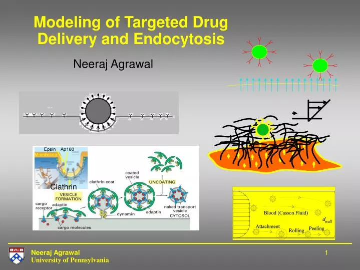
Clathrin. Modeling of Targeted Drug Delivery and Endocytosis. Neeraj Agrawal. Drug Carriers injected near the diseased cells Mostly drug carriers are in µm to nm scale Carriers functionalized with molecules specific to the receptors expressed on diseased cells

E N D
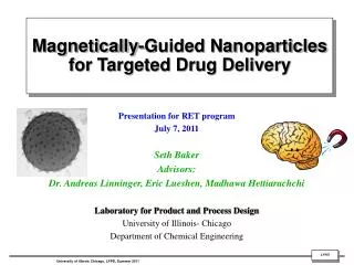
Clathrin Modeling of Targeted Drug Delivery and Endocytosis Neeraj Agrawal
Drug Carriers injected near the diseased cells Mostly drug carriers are in µm to nm scale Carriers functionalized with molecules specific to the receptors expressed on diseased cells Leads to very high specificity and low drug toxicity Targeted Drug Delivery
Motivation for Modeling Targeted Drug Delivery • Predict conditions of nanocarrier arrest on cell – binding mechanics, receptor/ligand diffusion, membrane deformation, and post-attachment convection-diffusion transport interactions • Determine optimal parameters for microcarrier design – nanocarrier size, ligand/receptor concentration, receptor-ligand interaction, lateral diffusion of ligands on microcarrier membrane and membrane stiffness
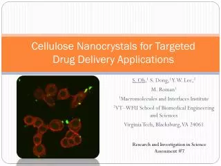
Cell 10-20 μm Antigen 20 nm Bead 100 nm Antibody 10 nm Glycocalyx 100 nm1,2,3 Glycocalyx Morphology and Length Scales Length Scales 1 Pries, A.R. et. al. Pflügers Arch-Eur J Physiol. 440:653-666, (2000). 2 Squire, J.M., et. al. J. of structural biology, 136, 239-255, (2001). 3 Vink, H. et. al., Am. J. Physiol. Heart Circ. Physiol. 278: H285-289, (2000).
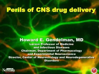
Effect of Glycocalyx (Experimental Data) Binding of carriers increases about 4 fold upon infusion of heparinase. Glycocalyx may shield beads from binding to ICAMs Mulivor, A.W.; Lipowsky, H.H. Am J Physiol Heart Circ Physiol 283: H1282-1291, 2002 Increased binding with increasing temperature can not be explained in an exothermic reaction In vitro experimental data from Dr. Muzykantov
Proposed Model for Glycocalyx Resistance • The glycocalyx resistance is a combination of • osmotic pressure (desolvation or squeezing out of water shells), • electrostatic repulsion • steric repulsion between the microcarrier and glycoprotein chains of the glycocalyx • entropic (restoring) forces due to confining or restricting the glycoprotein chains from accessing many conformations. S S=penetration depth
Parameter for Glycocalyx Resistance For a nanocarrier, k = 3.9*109 J/m4 Mulivor, A.W.; Lipowsky, H.H. Am J Physiol Heart Circ Physiol 283: H1282-1291, 2002
Simulation Protocol for Nanocarrier Binding Equilibrium binding simulated using Metropolis Monte Carlo. New conformations are generated from old ones by -- Translation and Rotation ofnanocarriers -- Translation of Antigens on endothelial cell surface Bond formation is considered as a probabilistic event Bell model is used to describe bond deformation =equilibrium bond length L=bond length Periodic boundary conditions along the cell and impenetrable boundaries perpendicular to cell are enforced
Monte-Carlo moves for bond-formation Select a nanocarrier at random. Check if it’s within bond-formation distance Select an antibody on this nanocarrier at random. Check if it’s within bond-formation distance. Select an antigen at random. Check if it’s within bond-formation distance. For the selected antigen, antibody; bond formation move is accepted with a probability If selected antigen, antibody are bonded with each other, then bond breakage move accepted with a probability
Binding Mechanics Multivalency: Number of antigens (or antibody) bound per nanocarrier Energy of binding: Characterizes equilibrium constant of the reaction in terms of nanobeads Radial distribution function of antigens:Indicates clustering of antigens in the vicinity of bound nanobeads These properties are calculated by averaging four different in silico experiments.
Effect of Antigen DiffusionIn silico experiments Increasing antigen concentration diminishes the effect of antigen diffusion.
Effect of Antigen FlexureIn silico experiments Allowing antigens to flex leads to higher multivalency.
Spatial Modulation of Antigens 500 nanocarriers (i.e. 813 nM) on a cell with antigen density of 2000/μm2 Nanobead length scale Diffusion of antigens leads to clustering of antigens near bound nanocarriers
Effect of GlycocalyxIn silico experiments Based on Glycocalyx spring constant = 1.6*10-7 N/m Presence of glycocalyx affects temperature dependence of equilibrium constant.
Antigen diffusion leads to higher nanocarrier binding affinity Diffusing antigens tend to cluster near the bound nanocarriers Glycocalyx represents a physical barrier to the binding of nanocarriers Presence of Glycocalyx not only reduces binding, but may also reverse the temperature dependence of binding Conclusions
Clathrin Multiscale Modeling of Protein-Mediated Membrane Dynamics:Integrating Cell Signaling with Trafficking Neeraj Agrawal
Endocytosis: The Internalization Machinery in Cells • Detailed molecular and physical mechanism of the process still evading. • Endocytosis is a highly orchestrated process involving a variety of proteins. • Attenuation of endocytosis leads to impaired deactivation of EGFR – linked to cancer • Membrane deformation and dynamics linked to nanocarrier adhesion to cells • Short-term Quantitative dynamic models for membrane invagination: Development of a multiscale approach to describe protein-membrane interaction at the mesoscale (m) • Long-term Integrating with signal transduction Minimal model for protein-membrane interaction in endocytosis on the mesoscale
Endocytosis of EGFR • A member of Receptor Tyrosine Kinase (RTK) family • Transmembrane protein • Modulates cellular signaling pathways – proliferation, differentiation, migration, altered metabolism • Multiple possible pathways of EGFR endocytosis – depends on ambient conditions • Clathrin Dependent Endocytosis • Clathrin Independent Endocytosis
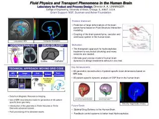
Membrane . epsin clathrin EGF epsin clathrin AP AP-2 - clathrin 2 epsin clathrin epsin epsin clathrin clathrin 2 - AP AP clathrin - 2 2 - AP Clathrin Dependent Endocytosis • One of the most common internalization pathway AP2 • Common theme: • Cargo Recognition – AP2 • Membrane bending proteins – Clathrin, epsin Clathrin polymerization Kirchhausen lab.
Trafficking Mechanism of EGFR Wiley, H.S., Trends in Cell biology, vol 13, 2003.
Overview Protein diffusion models Membrane models Model Integration Random walker Tale of three elastic models Preliminary Results
Linearized Elastic Model For Membrane: Monge-TDGL • Helfrich membrane energy accounts for membrane bending and membrane area extension. • In Monge notation, for small deformations, the membrane energy is z(x,y) Spontaneous curvature Bending modulus Frame tension Splay modulus • Consider only those deformations for which membrane topology remains same. • Force acting normal to the membrane surface (or in z-direction) drives membrane deformation The Monge gauge approximation makes the elastic model amenable to Cartesian coordinate system
Curvature-Inducing Protein Epsin Diffusion on the Membrane • Each epsin molecule induces a curvature field in the membrane KMC-move • Membrane in turn exerts a force on epsin Bound epsin position epsin(a) epsin(a+a0) where a0 is the lattice size, F is the force acting on epsin Metric Epsin performs a random walk on membrane surface with a membrane mediated force field, whose solution is propagated in time using the kinetic Monte Carlo algorithm
R Hybrid Multiscale Integration KMC TDGL #=1/De #=/t • Regime 1: Deborah number De<<1 or (a02/D)/(z2/M) << 1 • Regime 2: Deborah number De~1 or (a2/D)/(z2/M) ~ 1 Surface hopping switching probability Relationship Between Lattice & Continuum Scales Lattice continuum: Epsin diffusion changes C0(x,y) Continuum lattice: Membrane curvature introduces an energy landscape for epsin diffusion
Monge TDGL (linearized model) Radial distribution function Orientational correlation function Surface Evolution validation, computational advantage. Local TDGL vesicle formation. Integration with signaling Clathrin Dependent Endocytosis Interaction of Clathrin, AP2 and epsin with membrane Clathrin Independent Endocytosis Targeted Drug Delivery Interaction of Nanocarriers with fluctuating cell membrane. Applications
Local-TDGL(No Hydrodynamics) • A new formalism to minimize Helfrich energy. • No linearizing assumptions made. • Applicable even when membrane has overhangs • At each time step, local coordinate system is calculated for each grid point. • Monge-TDGL for each grid point w.r.to its local coordinates. • Rotate back each grid point to get overall membrane shape. Exact solution for infinite boundary conditions TDGL solutions for 1×1 µm2 fixed membrane
Potential of Mean Force • PMF is dictated by both energetic and entropic components x0 • Epsin experience repulsion due to energetic component when brought close. • Second variation of Monge Energy (~ spring constant). Test function • Non-zero H0 increases the stiffness of membrane lower thermal fluctuations Bound epsin experience entropic attraction.
Cell 10-20 μm Antigen 20 nm Bead 100 nm Antibody 10 nm Glycocalyx 100 nm1,2,3 Glycocalyx Morphology and Length Scales Length Scales 1 Pries, A.R. et. al. Pflügers Arch-Eur J Physiol. 440:653-666, (2000). 2 Squire, J.M., et. al. J. of structural biology, 136, 239-255, (2001). 3 Vink, H. et. al., Am. J. Physiol. Heart Circ. Physiol. 278: H285-289, (2000).