Download
1 / 21
230 likes | 404 Views
AGED-RELATED MACULAR DEGENERATION. (AMD). BY: Basiru Lee Leigh Mentor: Dr. Lee Angioletti Angioletti Retina Associates. The part of the eye dealing with AMD.

E N D
AGED-RELATED MACULAR DEGENERATION. (AMD) BY: Basiru Lee Leigh Mentor: Dr. Lee Angioletti Angioletti Retina Associates
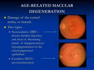
The part of the eye dealing with AMD. The RETINA: the Retina is a delicate lining in the back of the eye that creates impulses that travel through the optic nerve to the brain. The MACULA: the part of the eye that allows you to see fine details.
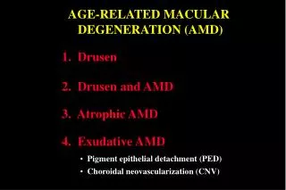
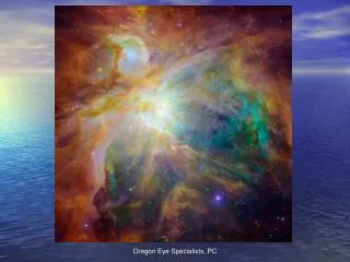
AMD is a disease associated with aging that gradually destroys the sharp central vision of the eye. AMD is also the leading cause of irreversible severe central visual loss. AMD mostly Affects adults 50 years and older in the United States. AMD affects the macula. AMD has two forms: Dry & Wet AMD. What is AMD
What are the causes and symptoms of AMD. • CAUSES: It can be cause by variety of factors like genetic, age, nutrition, smoking and sunlight exposure. • SYMPTOMS: 1. Difficulty in reading. 2.distorted vision. 3. loss of central vision. This symptom mostly happens in the Dry type.
Cures for Dry and Wet AMD. • Dry AMD has no treatment, but patients are told to take vitamins and see an ophthalmologist every now and then. • Wet AMD: 1.laser photocoagulation therapy. 2.photodynamic therapy (PDT). 3. injection of Anti-VEGF (Lucentis, Avastin, kenalog, etc)
It’s when cells and blood vessels beneath the macula break down and cause deposits in the eye. In the United States about 90% of the patients with AMD have the Dry type. It causes only 10% vision loss in the patients with Dry AMD. DRY AMD
WET AMD • Wet AMD happen when abnormal blood vessel growth forms beneath the Macula. • Only 10% of the patients with AMD have the Wet type in the Untied States. • It also causes 90% of the vision loss in the patients.
OBJECTIVE • To see if vitamins can reduce the risk of severe vision loss. • To combine two treatments for Wet AMD and see the results.
MATERIALS Laser (PDT) Fluorescein Angiography Kenalog Lucentis Amsler Grid Injection needle
PROCEDURE • DRY AMD. .Took pictures of their eyes. . They were told to take vitamins containing Zinc (80mg), copper (2mg), vitamins A,C and E. And to see the ophthalmologist on regular bases. • WET AMD. . Took pictures of the eye. . Dilate the eyes. . Then injected with Lucentis or Kenlog, followed up by PDT, or some times PDT by itself. .There eyes are later washed with an iodine solution. . Patients were told to come back every three months for more injection if the eye get worst.
PATIENT’S TREATMENT • This patients AMD wasn’t that severe so the patients was told to take small amounts of vitamins. • The patients also had to come to see the Doctor every six months.
PATIENTS TREATMENT. • The patient with the Wet AMD was severe and it was untreated for a while, the patient could have gone legally blind if the eye wasn’t treated, although the OD (Rigth eye) couldn’t see. • The patient was injected with Lucentis (0.5mg) and giving PDT. • He was released and told to come back in three month.
Expected Results • Dry AMD: Hope that the dry type doesn't develop into a wet type, or to lose most of his/her vision. Wet Type: We expected the Kenalog plus the PDT would lessen the amount swelling and coagulate abnormal blood vessels.
REFERENCES http://www.gene.com/gene/news//press- releases/display.do?method=detail&id=9147 http://www.nei.nih.gov/health/maculardegen/armd-facts.asp#1a http://www.macular-degenation.org/ Veteporfin in photodynamic therapy study group: by AM J> Ophthamol Hardy RA (2004). Retina. In D Vaughan et al., eds., General Ophthalmology, 16th ed., pp. 189–211. New York: Lange Medical Books/McGraw-Hill. http://www.stlukeseye.com/conditions/maculardegeration.asp. http://www.amd.org http://alconlabs.com/us/eo/conditions/AMD/causesAMD.jhtml
Acknowledgements • Dr. Lee Angioletti • Dr. sat • Harlem children society • MSKCC