Download
1 / 58
590 likes | 792 Views
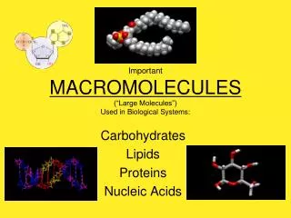
Biological Macromolecules. Chemistry and Chemical Biology Rutgers University. Overview. Bonds and Molecules Interactions in Biology Non-covalent: Hydrogen bonds, Hydrophobic interactions, Electrostatic interactions Covalent: Disulfide bonds, Coordinate bonds Proteins Nucleic Acids.

E N D
Biological Macromolecules Chemistry and Chemical Biology Rutgers University
Overview • Bonds and Molecules • Interactions in Biology • Non-covalent: • Hydrogen bonds, Hydrophobic interactions, Electrostatic interactions • Covalent: • Disulfide bonds, Coordinate bonds • Proteins • Nucleic Acids
Bonds and Molecules • Small Molecules and Macromolecules • Properties of Single Bonds (Covalent) • Chirality • Configuration • Conformation
Configuration b-D-glucopyranose a-D-glucopyranose • To go from a-D-glucose to b-D-glucose a bond has to be broken
L- and D- configurations of amino acids L-Threonine D-Threonine Mirror plane L-allo-Threonine D-allo-Threonine
Conformation • Described in terms of torsion angles • Rotation around bond • No bonds broken • Minimize non bonded interactions
Positive & Negative Torsion Angles • +Q: Clockwise rotation of front bond (about central bond) to eclipse the bond to the back • -Q:Counter clockwise rotation of front bond to eclipse the bond to the back • From http://www.currentprotocols.com/protocol/nca01c
Torsion Angle Nomenclature Saenger, Wolfram. Principles of Nucleic Acid Structure. Springer-Verlag New York Inc., 1984, p. 16.
Cyclohexanes Pyranose sugars Conformation Examples Voet, Donald and Judith G. Biochemistry. John Wiley & Sons, 1990, pp. 249-250.
Non-covalent Interactions in Biology • Non-covalent: • Hydrogen bonds • Hydrophobic interactions • Electrostatic interactions • Covalent: • Disulfide bonds • Coordinate bonds
Examples of Hydrogen Bonding Water Hydrogen Bonding Ice structure Voet, Donald and Judith G. Biochemistry. John Wiley & Sons, 1990, p. 30.
Hydrophobic interactions Clathrate hydrates Voet, Donald and Judith G. Biochemistry. John Wiley & Sons, 1990, p. 179.
Electrostatic interactions Example of electrostatic interaction in PDB entry 1HSA (MHC complex)
Disulfide Bridges Example of a disulfide bond in PDB entry 1HSA (MHC complex)
Coordinate Bonding N-ter Example of a Zinc Finger domain From PDB entry 1ZAA (ZIF268 protein) C-ter
Protein Building Blocks: Amino Acids • Amino acid sequence determines the 3D structure of a protein • 20 amino acids – modifications do occur post protein synthesis • L- Amino acids in normal proteins “corn crib” Voet, Donald and Judith G. Biochemistry. John Wiley & Sons, 1990, p. 68.
Amino Acids From www.bachem.com
Protein Building: Peptide Bonds • Individual amino acids form a polypeptide chain • The polypeptide chain is a component of a hierarchy for describing protein structure • The chain has its own set of attributes
S f y T f y f L y L f W y f y f w Q y T Conformation of the Polypeptide Chain • Omega (w): Rotation around the peptide bond Cn – N(n+1). It is planar and is 180 under ideal conditions • Phi (f): is the angle around N – Ca • Psi (y): is the angle around Ca – C • Values of f and y are constrained to certain values based on steric clashes of the R group. • Ramachandran plot: Defines characteristic patterns of torsion angles
Ramachandran Plot • Allowed & disallowed regions of f - y • Exceptions: • Gly has no limitation • Pro is constrained since its side chain binds back to the main chain • The F-y values for secondary structural elements are clustered • . Gray = allowed conformations. bA, antiparallel b sheet; bP, parallel b sheet; bT, twisted b sheet (parallel or anti-parallel); a, right-handed a helix; L, left-handed helix; 3, 310 helix; p,p helix.
Four Levels of Protein Structure • Primary, 1o • Amino acid sequence; Covalent bonds • Secondary, 2o • Local conformation of main-chain (polypeptide backbone) atoms (clustered F-Yangles); non-covalent interactions (H-bonds) TPEEKSAVTALWGKV
Four Levels of Protein Structure • Tertiary, 3o • 3D arrangement of all atoms in space (main-chain and side-chain); non-covalent interactions; covalent interactions may also contribute (e.g. S-S bonds) • Quaternary, 4o • Interaction of subunit chains; non-covalent interactions b2 a2 a1 b1 b2
Secondary Structure: Alpha Helices • If N-terminus is at bottom, then all peptide N-H bonds point “down” and all peptide C=O bonds point “up”. • C=O(i) is H-bonded to N-H(i+4). • Features: • 3.6 residues per turn • Rise/residue = 1.5 Å • Rise/turn = 5.4Å
a Helix • R-groups in a-helices: • extend radially from the core, • shown in helical wheel diagram. • Can have varied distributions Polar Hydrophobic Amphipathic
Secondary Structure: b Sheet • Stabilized by H-bonds between N-H & C=O from adjacent stretches of strands • Peptide chains are fully extended pleated shape because adjacent peptides groups can’t be coplanar.
Beta Sheets Parallel beta sheet Not optimum H-bonds; less stable Antiparallel beta sheet Optimum H-bonds; more stable
The Beta Turn AA2 AA3 • H-bond between N-H(i) and C=O(i+3) • Beta-turns can have 2 different conformations AA1 AA4
Motifs • Motif (structural motif) : Arrangement of secondary structural elements • May not have same/similar biochemical function • May also be called super-secondary structure • Note: Sequence motifs are recognizable aa sequence with a biochemical function Helix-turn-helix motif PDB ID 1lmb Greek-key motif PDB ID 3ix0 b-a-b motif PDB ID 2gcf
Examples of Tertiary Interactions Charge based interactions 62R:163E 55E:170R Hydrophobic interactions 189V 201L 213I 215L 266L Disulfide bond 203C:259C domains 1 2 3
Domains • Collection of several secondary structural elements and/or motifs • Tertiary structure • Usually has a hydrophobic core • May be a complete protein or part of a protein • May be stabilized by covalent interactions (S-S bonds, coordinate covalent bonds) • Types: alpha, beta, alpha/beta (Levitt-Chothia classification)
Protein Domain Examples Globin fold (Myoglobin) PDB ID 1ajg TIM Barrel PDB ID 1tim Jelly roll PDB ID 1k5j
Domain Classifications • Sequence based • PFAM: protein families, represented by multiple sequence alignments • Structure based • SCOP: Structural Classification of Proteins • CATH: Class(C), Architecture(A), Topology(T), Homologous superfamily (H)
Class(C)derived from secondary structure content is assigned automatically • Architecture(A)describes the gross orientation of secondary structures, independent of connectivity. • Topology(T) clusters structures according to their topological connections and numbers of secondary structures [ http://www.biochem.ucl.ac.uk/bsm/cath_new/ ]
Quaternary Structure • Composed of 2 or more polypeptide chains • Intermolecular interactions may be non-covalent or covalent • Inappropriate interactions in disease (sickle cell hemoglobin mutant E6V in b2)
References • "Introduction to protein structure", Brandon and Tooze, 3, 21, 1999. • Voet, Donald and Judith G. Biochemistry. John Wiley & Sons, 1990 • “Protein Structure and Function”, Petsko and Ringe, New Science Press Ltd., 2004
Nucleic Acid Structures Base Sugar Phosphate
Building Blocks in Nucleotides Bases Purines and Pyrimidines Bases in DNA (dA, dT, dG, dC) Bases in RNA (A, U, G, C) Sugars DNA has deoxy ribose sugar RNA has ribose sugar Phosphate deoxy-ribose ribose Bases C5’ C5’ C5’ P Phosphate Sugars
Watson Crick Base Pairs Geometry of A:T and G:C base pairs are isosteric
Tautomeric structures • Keto vs enol • Different hydrogen bonding patterns
Base Morphology: Base pair twist • Looking down helix axis • 36 degree base pair twist in B-DNA B-DNA structure in 424D
Anti vs. syn Syn conformation Anti conformation