Download
1 / 19
200 likes | 516 Views
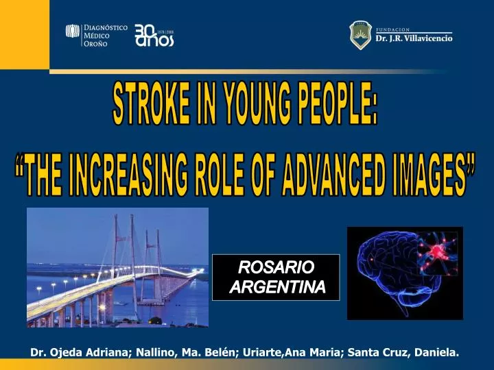
STROKE IN YOUNG PEOPLE: “THE INCREASING ROLE OF ADVANCED IMAGES”. ROSARIO ARGENTINA. Dr. Ojeda Adriana; Nallino, Ma. Belén; Uriarte,Ana Maria; Santa Cruz, Daniela. INTRODUCTION. STROKE is a leading cause of death and disability in developed countries.

E N D
STROKE IN YOUNG PEOPLE: “THE INCREASING ROLE OF ADVANCED IMAGES” ROSARIO ARGENTINA Dr. Ojeda Adriana; Nallino, Ma. Belén; Uriarte,Ana Maria; Santa Cruz, Daniela.
INTRODUCTION STROKE is a leading cause of death and disability in developed countries. Ischemic stroke in young adults ( 15-45 ys) is an unexpected event . Etiological spectrum is varied and differs significantly from the underlying causes in older patients.
PURPOSE To highlight the increasing role of neuroimaging in the diagnosis, prognosis and treatment in young stroke patients. The most frequent causes of ischemic stroke are considered and the corresponding images are described.
MATERIALS AND METHODS • 30 patients (mean age 35 ys.) with Acute Ischemic Stroke diagnosis were retrospectively included and analized at our institution over one year period. • Neurovascular imaging • MSCT 8 - 64 channels: Unenhanced CT, CTA, CT PERFUSION • MRI 1.5 T: Conventional MRI, MRA, DWI, PWI • CLINICAL DATA-RISK FACTORS-CARDIOVASCULAR EXAMS-LAB TEST • .
STROKE TOAST CLASSIFICACION
ATHEROSCLEROSIS: • Dislipidemy • Hipertension + smoking CARDIOEMBOLISM: • 2 P.F.O. • 1 Atrial Septal Aneurysm UNDETERMINED: • 3 Complete evaluation • 1 Incomplete • DISSECTION: • 5 Vertebral A • 1 Basilar A • 2 Carotid A MISCELLANEOUS: • 3 Migrainous infarction • 2 moya-moya • 2 cerebral venous thrombosis • 2 systemic vasculitis • 1 SLE • 1 Antiphospholipid syndrome • 1 vasoespasm secondary
COMPLETE WORK-UP INCLUDES • CT or MRI scanning, CTA or MRA. • Echocardiography • Routine and Specific Lab Test • Risk Factors analysis • Drug abuse report
T2 DWI Healthy 40 year-old. female patient referring acute headache, neck pain, dizziness and vomits T1 LEFT VERTEBRAL DISSECTION
45 year-old male patient with severe trauma history due to a car accident 15 days before showing sudden onset aphasy INTIMAL FLAP DOUBLE LUMEN
CAROTID DISSECTION
FLAIR DWI ADC 24 year-old female patient with language and speech disorder (aphasia) ANTIPHOSPHOLIPID SYNDROME
DWI 30 year-old male patient with drop attack, vomits and stupor during a migraine episode. FLAIR T2 DWI MIGRAINOUS INFARCTION
45 year old female patient with right hemiparesis and aphasia. Unenhanced CT normal CBV CBF MTT PERFUSION CT CRYPTOGENIC STROKE
DISCUSSION The etiologic spectrum is quite broad, thus the investigation represents a challenging diagnosis. Atherosclerotic arteriopathy was a rare cause of stroke. Artery dissection was the most frequent cause identified in our series, as has already been reported in the medical literature. Underterminaded cause represented a minor percentage compared to other studies.
DISCUSSION In our study the sample is relatively small and show selection bias. Our results emphasize the importance of a standardized protocol and a comprehensive evaluation of stroke in young patients. To carry out multicentrical randomized studies to assay the stroke incidence in Argentinean young people.
CONCLUSION • The aims of NEUROIMAGING in the study of young stroke patients are: • to confirm the ischemic nature of the lesion • to determine its location • to verify the patency of major neck and intracranial arteries • MRI and MSCT offer great sensitivity and specificity
BIBLIOGRAFÍA • Heather Bevan, MD, Khema Sharma, MD, and Walter Bradley, MD. Stroke in young adults. Stroke 1990; 21:382-386. • Jose Ibiapina Siqueira MD, PhD; Santos Antonio Carlos MD; Fabio, Soraria Ramos Cabete MD, Sakamoto, Americo C. MD, PhD. Cerebral infarction in patients aged 15 to 40 years, Stroke 1996; 26:2016-2019 • Kristensen, Bo MD; Malm, Jan PhD; Carlberg, Bo PhD; Stegmayr, Birgitta PhD. Epidemiology and Etiology of Ischemic Stroke in Young Adults Aged 18 to 44 Years in Northern Sweden. Stroke 1997; 28:1702-1709. • J.F. Varona, J.M Guerra, F. Bermejo, J.A. Molina, A. Gomez. Causes of ischemic Stroke in Young Adults, and Evolution of the Etiological Diagnosis over the Long Term. European Neurology 2007; 57:210-218. • Fahmi Yousef Khan, risk factors of young ischemic stroke in Qatar, Clinical Neurology and Neurosurgey 2007; 109:770-773. • Michael T.Y. Chan, Zurab G. Nadareishvili, John W. Norris. Diagnostic Strategies in Young Patients with Ischemic Stroke in Canada. Can. J. Neurol. Sci. 2000; 27:120-124. • Tali Jonas Kimchi, MD, Ronit Agid, MD, Seon-Kyu Lee, MD, Karel G. Ter Brugge, MD. Arterial Ischemic Stroke in Children. Neuroimag. Clin. N. Am. 2007; 17:175-187. • Ashok Srinivasan, MD, Moyank Goyal, MD, Faisal Al Azri, MD, Cheemun Lum, MD. State-of-the-Art Imaging of Acute Stroke. Radiographics 2006; 26: S75-S95. • C. Uggetti. Stroke in young people:imaging. Neurol. Sci. 2003; 24:S15-S16. • Christine M. Flis, H. Rolf Jâger, Paul S. Sidhu. Carotid and vertebral dissections: clinical aspects, imaging features and endovascular treatment. Eur. Radiol. 2007; 17: 820-834. • Mathieu H. Rodallec, MD, Veronique Marteau, MD, Sophie Gerber, MD, Loîc Desmonttes, MD, Marc Zins, MD. Craniocervical Arterial Dissection : Spectrum of Imaging Findings and Differential Diagnosis. Radiographics 2008; 28: 1711-1728. • A.T. Vertinsky, N.E. Schwartz, N.J. Fischbein, J. Rosenberg, G.W. Albers, G. Zaharchuk. Comparison of Multidetector CT Angiography and MR Imaging of Cervical Artery Dissection. AJNR 2008; 29: 1753-60. • Marie-Germaine Bousser, KMichael A Welch. Relation between migraine and stroke. Lancet Neurol 2005; 4:533-42. • Kurth T, Gaziano JM, Cook NR et al. Migraine and risk of cardiovascular disease in women. JAMA 2006; 296
MUCHAS GRACIAS! THANKS! Rosario Argentina