Download
1 / 17
170 likes | 466 Views
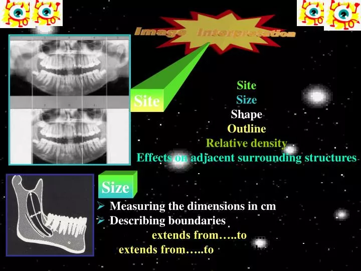
Site. Size. Measuring the dimensions in cm Describing boundaries (the lesion extends from…..to …..in one dimension and extends from…..to ……in the other dimension). Site Size Shape Outline Relative density Effects on adjacent surrounding structures. Shape. Monolocuar.

E N D
Site Size • Measuring the dimensions in cm • Describing boundaries • (the lesion extends from…..to…..in one dimension • and extends from…..to……in the other dimension) Site Size Shape Outline Relative density Effects on adjacent surrounding structures
Shape Monolocuar • Monolocular/unilocular • Multilocular • Pseudoloculated • Round • Oval • Scalloped/undulating • Irregular Pseudolocuar Multilocuar
Outline *Well-defined • Smooth • Punched-out • Corticated: a thick or thin surrounding RO line • Sclecotic: a non-uniform RO boundary • Oval • Encapsulated • Irregular Well-defined with a corticated margin Well-defined without a corticated margin Poorly-defined *Poorly-defined
Effects on adjacent surrounding structures Relative density • Bony expansion 2. Displacement ID canal 3. Thinning of cortex • Uniformly RL • RL with RO (mixed) • RO • Tooth displacement
www.dent.ucla.edu/sod/depts/oral_rad/courses/DS422b/ Question: Please describe the lesion as indicated by yellowish arrow There is a well-defined unilocular round shaped circumcoronal radiolucence with corticated margin over the submerged tooth 38 extending from left retromolar area down to the mandibular angle and from distal aspect of tooth 37 up to two/thirds of left ramus area, measuring approximately 3 5 cm in diameter.
A sequential approach to radiological interpretation Panoramic radiography revealed the patient to be fully edentulous. All bony outlines were within the normal range except for a 4.5 x 3.0 cm well-demarcated, unilocular homogeneous radiolucency with smooth well-corticated outline in the left body of the mandible. The lesion extended from the premolar region back to 1.5 cm anterior to the posterior margin of the mandibular ramus. There was slight expansion of the cortical outline of the lower border in the left antigonial notch region.
A sequential approach to radiological interpretation The adjacent mandibular canal was inferiorly displaced. Canal cortical outlines were intact with no evidence of resorption and the paranasal sinuses were clear. A root fragment was noted in the region of the radiolucency in the left mandible, and there were several areas of the well- delineated radiolucency in the left mandible was that of a benign cyst or tumor.
A sequential approach to radiological interpretation Panoramic radiography alsorevealed a well-delineated radiolucency rimmed by an ovoid 3.5 x 2.5 cm calcified margin, superimposed over the left mandibular ramus. The radiographic shadow of the calcified soft tissue lesion extended superiorly to the level of the mandibular sigmoid notch and 1.5 cm below the head of the left mandibular condyle, and inferiorly to 1 cm below the left mandibular foramen and lingula
A sequential approach to radiological interpretation The principal differential interpretations were carotid aneurysm and calcified lymph node. Although carotid bruit was not clinically detected, the risk of a carotid aneurysm mandated prompt investigation of this radiographic finding. To elucidate further the position of this calcified soft-tissue lesion and the boundaries of the mandibular radiolucency, an axial CT examination was performed.
Axial CT Axial CT Axial CT Maxilla 1st cervical vertebra A sequential approach to radiological interpretation The CT confirmed the presence of the calcified-rimmed soft tissue ventral and lateral of the first cervical vertebral body and skull base. This was interpreted as compatible with aneurysm or psedoaneurysmal dilation of the internal carotid artery, measuring as large as 2.4 cm. Degenerative changes in the cervical spine were noted.
Axial CT Axial CT Mandible, lower portion Mandible, upper portion A sequential approach to radiological interpretation Lower CT slices through the body of the mandible confirmed the homogeneously radiolucent cystic lesion with a benign appearance. There was evidence of buccal and lingual cortical expansion with attenuation. In view of the report of a probable carotid aneurysm, CT angiography was prescribed to relate this lesion to its surrounding structures.
A sequential approach to radiological interpretation CT angiography revealed the calcified mass was intimately related to tortuous internal and external carotid arteries. Careful reformatting at various angulations failed to demonstrate a direct continuity between the internal carotid and the presumed aneurysm; however due to structural superimposition CT failed to provide a definite answer. MRI was selected to elucidate further structures obscured in the CT angiograms.
A sequential approach to radiological interpretation MRI revealed bright signals for the carotid artery and jugular veins bilaterally, but failed to demonstrate an aneurysm. The contents of the lesion in the left mandibular body had intermediate signal intensity
A sequential approach to radiological interpretation Angio MaxIP MRI revealed the carotids were found to be tortuous. There was no evidence of an aneurysm of the left internal carotid artier. The surgeon wanted additional verification of the absence of a carotid aneurysm and ordered ultrasonography.
A sequential approach to radiological interpretation Ultrasound images showed no evidence of aneurysm. Both carotid bifurcations were tortuous. There was evidence of atherosclerotic plaque in the left and right carotid bifurcation bulbs and the proximal region of the left internal carotid artery. The systolic velocity ratio of right internal to common carotid artery was 0.59 whereas the diastolic velocity ratio was 0.78; for the left side the respective ratios were 0.96 and 1.47. These ratios are within the normal range.
A sequential approach to radiological interpretation Final Diagnosis Mandibular lesion Glandular odontogenic cyst Neck lesion The calcified lesion in the soft tissues adjacent to the first cervical vertebra and extending to the carotid space was not a carotid aneurysm, but rather a calcifying ‘cystic’ mass probably representing a lymph node.