Download
1 / 77
780 likes | 1.11k Views
Introduction to Clinical Electrocardiography. Gari Clifford, PhD Andrew Reisner, MD Roger Mark, MD PhD. Electrocardiography. The heart is an electrical organ, and its activity can be measured non-invasively Wealth of information related to: The electrical patterns proper

E N D
Introduction to Clinical Electrocardiography Gari Clifford, PhD Andrew Reisner, MD Roger Mark, MD PhD
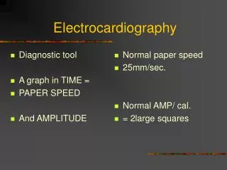
Electrocardiography • The heart is an electrical organ, and its activity can be measured non-invasively • Wealth of information related to: • The electrical patterns proper • The geometry of the heart tissue • The metabolic state of the heart • Standard tool used in a wide-range of medical evaluations
A heart • Blood circulates, passing near every cell in the body, driven by this pump • …actually, two pumps… • Atria = turbochargers • Myocardium = muscle • Mechanical systole • Electrical systole
To understand the ECG: • Electrophysiology of a single cell • How a wave of electrical current propagates through myocardium • Specific structures of the heart through which the electrical wave travels • How that leads to a measurable signal on the surface of the body
Once upon a time, there was a cell: K+ K+ 2 K+ 3 Na+ ATPase
a myocyte time Intracellular millivoltage Resting comfortably -90
Depolarizing trigger time Intracellular millivoltage
Na channels open, briefly time Intracellular millivoltage
Mystery current time Intracellular millivoltage In: Na+
Ca++ is in balance with K+ out time Intracellular millivoltage In: Na+
Excitation/Contraction Coupling: Ca++ causes the Troponin Complex (C, I & T) to release inhibition of Actin & Myosin time Intracellular millivoltage In: Na+
More K+ out; Ca++ flow halts Ca++ in; K+ out time Intracellular millivoltage In: Na+
Sodium channels reset In: Ca++; Out: K+ time Intracellular millivoltage In: Na+ Out: K+
Higher resting potential Few sodium channels reset Slower upstroke time Intracellular millivoltage In: Na+
Slow current of Na+ in; note the resting potential is less negative in a pacemaker cell a pacemaker cell time Intracellular millivoltage -55
Threshold voltage a pacemaker cell time Intracellular millivoltage -40
Ca++ flows in time Intracellular millivoltage
. . . and K+ flows out time Intracellular millivoltage
. . . and when it is negative again, a few Na+ channels open time Intracellular millivoltage
How a wave of electrical current propagates through myocardium • Typically, an impulse originating anywhere in the myocardium will propagate throughout the heart • Cells communicate electrically via “gap junctions” • Behaves as a “syncytium” • Think of the “wave” at a football game!
The dipole field due to current flow in a myocardial cell at the advancing front of depolarization. Vm is the transmembrane potential.
Important specific structures • Sino-atrial node = pacemaker (usually) • Atria • After electrical excitation: contraction • Atrioventricular node (a tactical pause) • Ventricular conducting fibers (freeways) • Ventricular myocardium (surface roads) • After electrical excitation: contraction
The Idealized Spherical Torso with the Centrally Located Cardiac Source (Simple dipole model)
Clinical Lead Placement • Einthoven Limb Leads:
The temporal pattern of the heart vector combined with the geometry of the standard frontal plane limb leads.
Vagal stimulation makes the resting potential MORE NEGATIVE. . . Neurohumeral factors time Intracellular millivoltage
Neurohumeral factors time Intracellular millivoltage . . . and the pacemaker current SLOWER. . .
. . . and raise the THRESHOLD time Intracellular millivoltage
Catecholamines make the resting potential MORE EXCITED. . . time Intracellular millivoltage
. . . and speed the PACEMAKER CURRENT. . . time Intracellular millivoltage
. . . and lower the THRESHOLD FOR DISCHARGE. . . time Intracellular millivoltage
time Intracellular millivoltage Vagal Stimulation:
time Intracellular millivoltage Adrenergic Stim.
Arrhythmias - Not firing when you should - Firing when you shouldn't - All of the above (Reentrance)
Firing when you shouldn't • Usually just a spark; rarely sufficient for an explosion • “Leakiness” leads to pacemaker-like current • Early after-depolarization • Late after-depolarization