Download
1 / 48
480 likes | 599 Views
Τεχνική ενδοφλεβικού αποκλεισμού υπερηχογραφικά υποβοηθούμενη. Χ. Ν. Μπακογιάννης Λέκτορας Αγγειοχειρουργικής Α’ Χειρουργική Κλινική ΕΚΠΑ Γ.Ν.Α. ¨Λαϊκό¨. ve. RFA Technology.

E N D
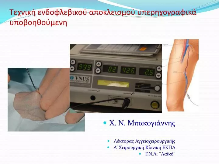
Τεχνική ενδοφλεβικού αποκλεισμού υπερηχογραφικά υποβοηθούμενη • Χ. Ν. Μπακογιάννης • Λέκτορας Αγγειοχειρουργικής • Α’ Χειρουργική Κλινική ΕΚΠΑ • Γ.Ν.Α. ¨Λαϊκό¨
RFA Technology In the original radiofrequency catheter system, the catheter was pulled through the vein while feedback is controlled with a thermocouple to a temperature of 85°C to avoid thermal injury to the surrounding tissues or carbonization of the vein wall. With the new system, the catheter is held in place while energy heats the catheter to a specified temperature of 120ºC. As the vein is denatured by heat, it contracts around the catheter
Technique- Procedure Duplex ultrasonographyis used to confirm and map all areas of reflux and to trace the path of the refluxing great saphenous trunk from the saphenofemoral junction down the leg to the lower thigh or upper part of the calf. The vein, the saphenofemoral junction, and the anticipated entry point are marked in some way on the skin The Seldinger technique is used to place a guidewire into the vessel, and an introducer sheath is passed over the guidewire, which is removed. The ClosureFast catheter is passed through the sheath, and the tip is advanced to 2 cm below the saphenofemoral junction under duplex ultrasonographic visualization. The catheter must not extend into the femoral vein because injury to the femoral vein may cause deep vein thrombosis.
Technique- Procedure With ultrasonographic guidance, a local anesthetic agent is injected into the tissues surrounding the great saphenous vein above and within its fascial sheath. The anesthetic is injected along the entire course of the vein from the catheter insertion point to the saphenofemoral junction. In most patients, 200-400 mL of lidocaine 0.1% is sufficient to both anesthetize and compress the vessel. Note the importance of delivering the anesthetic agent in the correct intrafascial location, with a volume sufficient to compress the vein and dissect it away from other structures, such as nerves, along its entire length. the previous system, after the temperature reaches 85°C and remains constant for 15 seconds, the catheter tip is slowly withdrawn at a rate of approximately 1 cm per minute (1 mm every 6 seconds). In the new system, two 20-second cycles are performed in the proximal section, after which the catheter is withdrawn 7 cm as per catheter markings. The next 20-second cycle is repeated once, and, if 120ºC is maintained, the catheter is then withdrawn another 7 cm until the entire vein is treated.
Histologic Findings Immediately after treatment, biopsy specimens show a significant reduction in the size of the vein lumen, with denudation of endothelium, thrombus formation, thickened vessel walls, loss of collagen birefringence, and inflammatory changes. The zone of thermal damage is limited to 2 mm beyond the point of contact with the electrodes. In more than 90% of patients, biopsy specimens demonstrate complete occlusion of the vein lumen 6 weeks after treatment. The lumen is completely ablated in most areas, with some portions of the vessel demonstrating a small residual lumen containing organized fibrous thrombi. Birefringence is present, and new collagen growth is evident
Cost-effectiveness of treatment In a small randomizedstudy of 28 patients, a basic cost-analysis demonstrated thatVNUS Closure was more expensive than conventional surgery interms of direct costs, but the costs to society were significantlylower in view of the rapid return to work in the RFA group. RFAperformed in an office setting may be cheaper than traditionalsurgery the cost of procedures will be heavily influencedby clinician preferences on type of anaesthetic, the use ofconcomitant ambulatory phlebectomy and whether to perform bilateralprocedures in a single sitting, thus saving the cost of an additionalRFA catheter
Τεχνική ενδοφλεβικού αποκλεισμού υπερηχογραφικά υποβοηθούμενη Μεταπτυχιακό Προγραμμα Ιατρικής Σχολής ΕΚΠΑ: “Ενδαγγειακές Τεχνικές”